

Anatomy of the female pelvis - Atlas of the human body using cross-sectional imaging
Anatomy of the female pelvis - Atlas of the human body using cross-sectional imaging
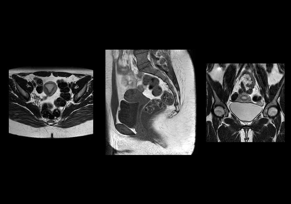
Magnetic resonance (MR) imaging is a valuable technique for the non-invasive evaluation of the female pelvic region (for example diagnosing or staging developmental anomalies, leiomyomas, adenomyosis, vaginal neoplasms, endometrial or cervical carcinoma, ovarian cysts, endometriosis, teratomas, polycystic ovaries or other ovarian masses), but also for the study of the normal anatomy of the female pelvis.
In this anatomy study, the user has access to a user-friendly and dynamic tool to learn and understand the main anatomical structures of the female pelvis.
Cross-sectional anatomy: Magnetic Resonance Imaging (MRI) of the female pelvic region
An MR was performed on a healthy woman with two kinds of weightings: axial, coronal and sagittal spin-echo T2-weighted MR images and axial spin-echo gadolinium-enhanced fat-saturated T1-weighted MR images.
DICOM images (from our PACS (Picture Archiving and Communicating System)) were exported to JPEG format and resized with Adobe Photoshop.
Interactive anatomical features have been developed with Adobe Animate.
This anatomical atlas was especially designed for radiologists, gynecologists, medicine students and residents.
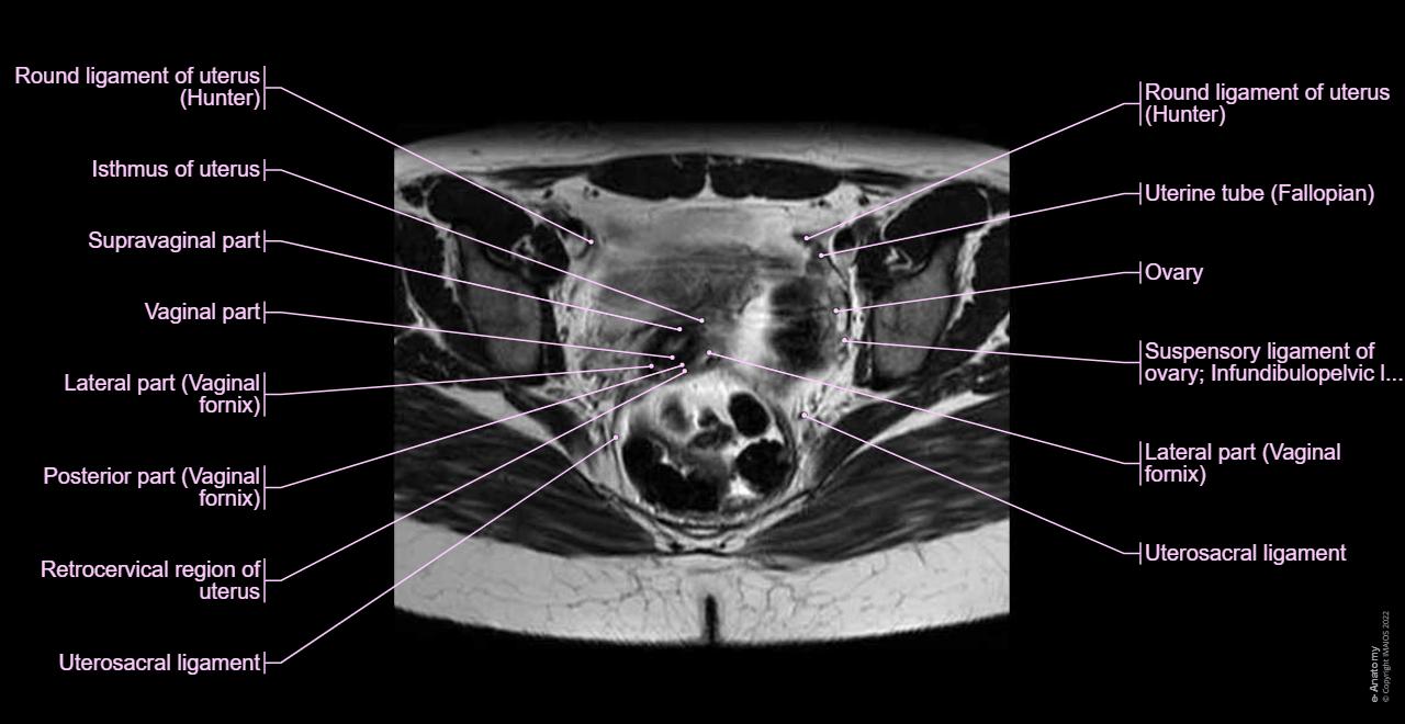
Anatomy of the human female pelvis: how to use the anatomical labels
On "Anatomical parts", the user can choose the type of anatomical labels he wishes to display on the exam:
- Genital organs: uterus (with uterine zonal anatomy: endometrium, myometrium, junctional zone), cervix (central zone, cervical stroma), parametria, vagina, ovaries and adnexa with follicles
- Urinary system including urinary bladder, ureter, urethra
- Digestive system including rectum and sigmoid colon
- Pelvic diaphragm
- Pelvic girdle
- Spaces and foramens of the pelvic region
- Muscles
- Arteries
- Veins
- Nerves
- Pelvic measurements
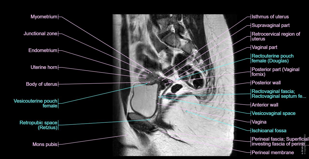
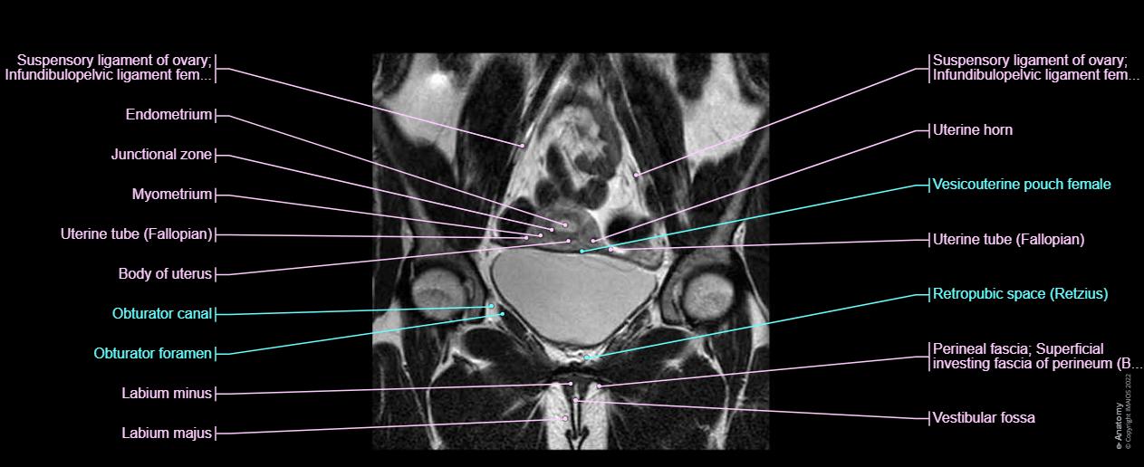
The user is able to choose between one of the three planes available (axial, coronal or sagittal) through the vertical menu on the left, which also provides cross-references.
By moving the mouse cursor over a particular anatomical area of the pelvic region, this region is highlighted and the labels are displayed: peritoneal cavity, subperitoneal space and perineal region.
No content available
