

Anatomy of the ankle and foot (MRI) - atlas of the human body using cross-sectional imaging
Anatomy of the ankle and foot (MRI) - atlas of the human body using cross-sectional imaging
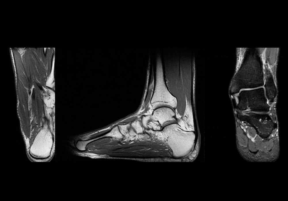
This cross-sectional human anatomy atlas of the ankle and foot is a new tool based on MR images of the human body.
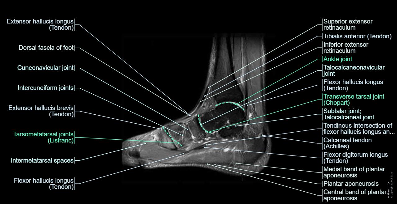
Anatomical structures of the ankle and foot and specific regions (major joints) are visible as dynamic labeled images.
Cross-sectional anatomy: MRI of the ankle and feet
A magnetic resonance imaging (MRI) was performed on a normal subject; with spin-echo T1 weighted images and spin-echo fat-saturated proton density weighted images (3 usual planes used for osteo-articular imaging: axial, coronal, and sagittal).
From our PACS (Picture Archiving and Communicating System), data and DICOM images were exported as JPEG images. Minor changes were done with Adobe Photoshop, and then, the images were interactively labeled using Adobe Animate.
This module is a comprehensive and affordable learning tool for medical students and residents and especially for physicians, anatomists, rheumatologists, orthopedic surgeons and radiologists. It is also a useful communication tool to teach patients anatomy and pathology.
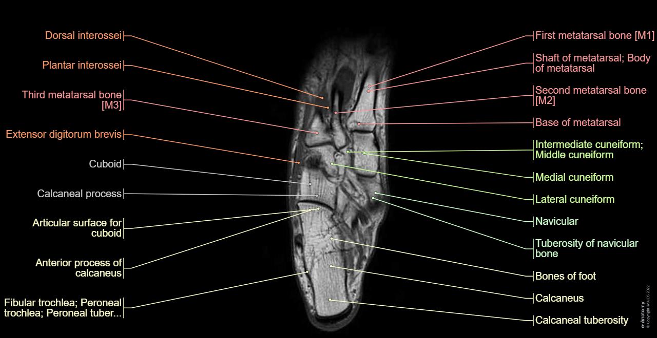
It provides access to an atlas and to images in the axial planes, allowing the user to learn and review orthopedic anatomy interactively. All images were labeled, providing an invaluable teaching resource.
We used the Terminologia Anatomica to create the anatomical labels. This terminology is the international standard on human anatomy (it supersedes the previous standard, Nomina Anatomica since 1998). It was developed by the Federative Committee on Anatomical Terminology (FCAT) and the International Federation of Associations of Anatomists (IFAA).
Anatomy of the lower limb (hip, thigh, knee, leg, ankle and foot): how to view the anatomical labels
On "Anatomical parts" the user can access the groups of anatomical structures:
- Bones
- Joints
- Ligaments
- Muscles
- Tendons
- Fascia
- Arteries
- Veins
- Nerves
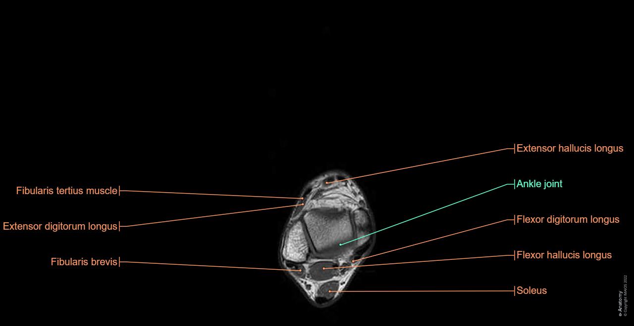
By moving the mouse cursor over a specific area, this region is highlighted and its labels displayed: anterior and posterior region of the leg, posterior and anterior ankle region, metatarsal region, plantar region.
The vertical menu on the left provides cross-references and a 3D medical illustration of the foot skeleton.
Tear lines represent the main joints of ankle and foot:
- Ankle joint
- Transverse tarsal joint
- Tarsometatarsal joints
The "Quiz mode" allows the user to self-evaluate his study progress.
No content available
