

Anatomy of the autonomic nervous system
Anatomy of the autonomic nervous system
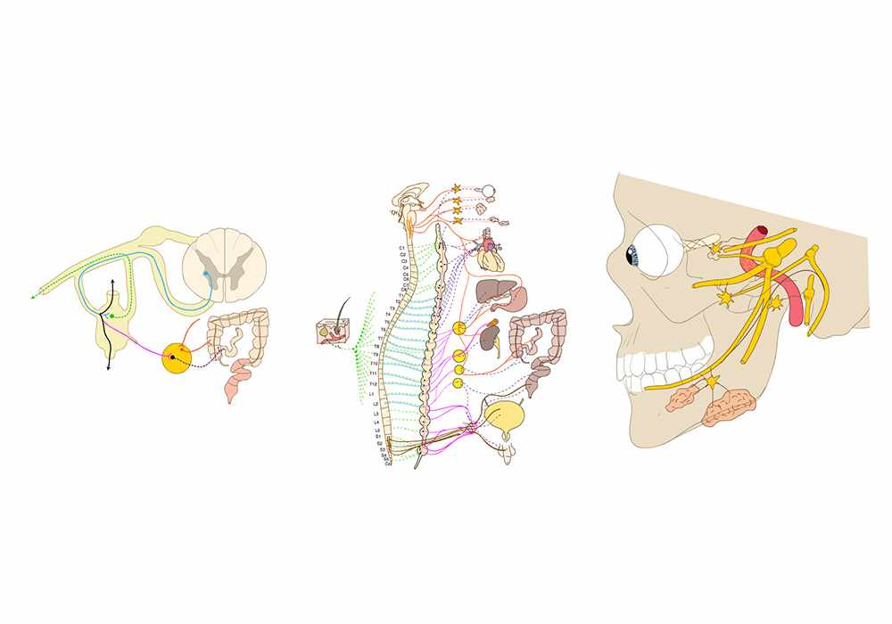
Three anatomical diagrams are represented separately in this module. In fact, the study of the autonomic nervous system (sympathetic and parasympathetic) is often based on the study of its organs.
The three illustrations of this module are original work designed by MD Micheau, Antoine and summarize diagrams of the functioning of the autonomic nervous system.
Diagrams of the autonomic nervous system
These human anatomy vector diagrams were made using Adobe Illustrator and then integrated into the modules using Adobe Animate.
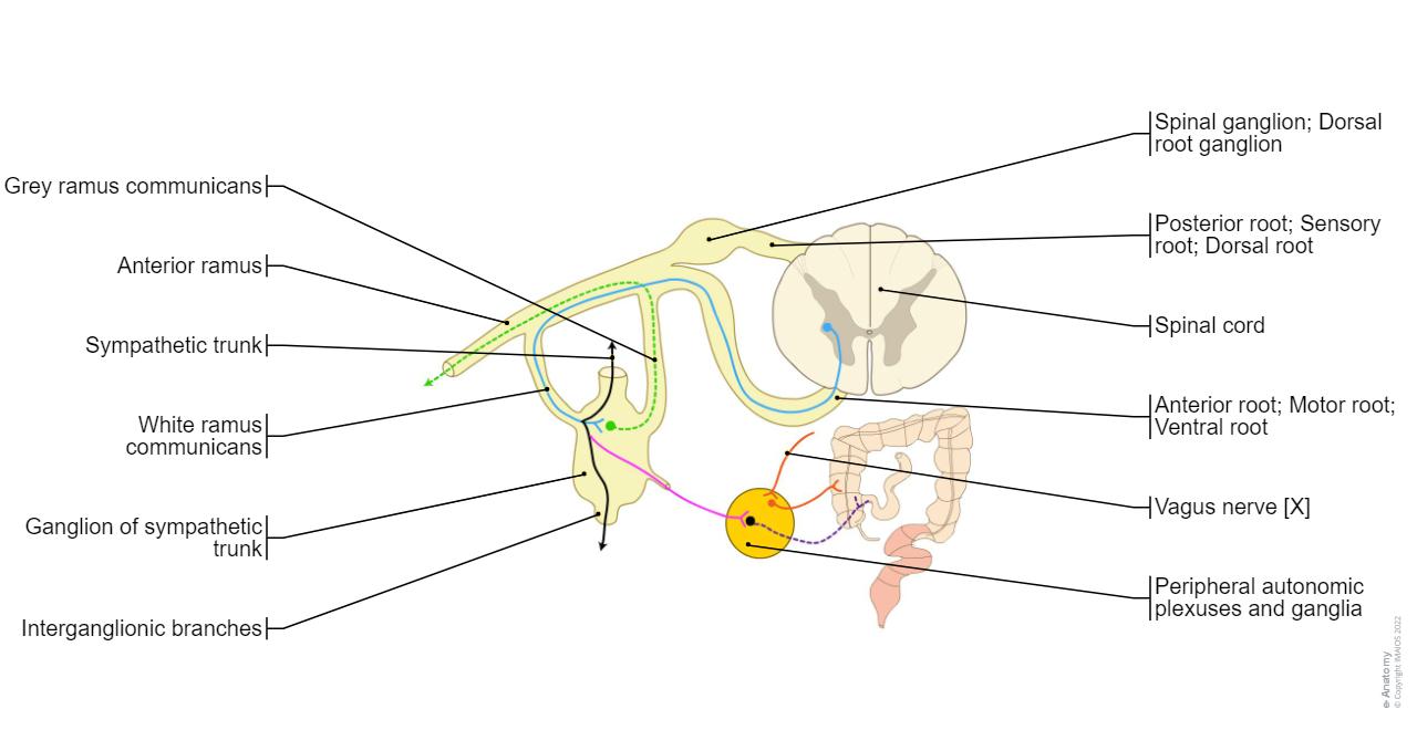
- The first diagram is a section of the autonomic nervous system, with the fibres issuing from the spinal nerve (white and grey communicating branch), the relations with the sympathetic trunk ganglia then with the visceral plexuses and visceral ganglia.
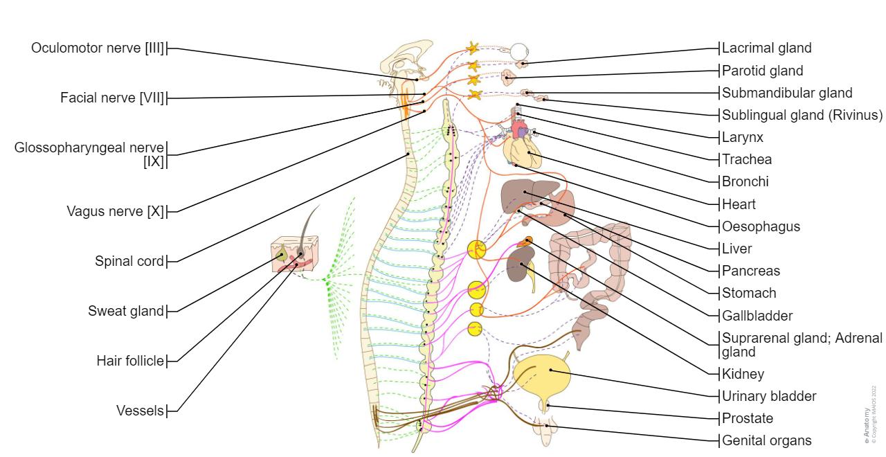
- The second diagram brings together all the types of autonomic innervation of the face, thorax and abdominal-pelvic organs while detailing the ganglia (cervical, thoracic, lumbar and sacral) of the sympathetic trunk.
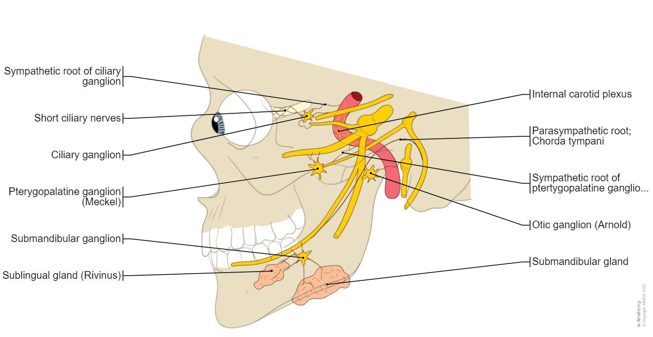
- The latter summarises the different sympathetic and parasympathetic structures of the head and face, (ciliary ganglion, pterygopalatine ganglion, otic ganglion) as well as the branches communicating with the cranial nerves
No content available
