

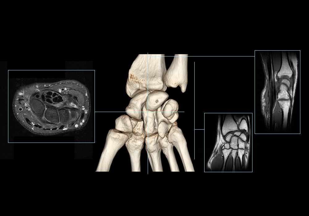
Anatomy of the wrist (cross-sectional imaging on 3T MR and 3D medical pictures)
Anatomy of the wrist (cross-sectional imaging on 3T MR and 3D medical pictures)
Images provided by Sorin Ghiea & Emi Preda - MD
On this module on the anatomy of the wrist in 3T MRI, we labeled over 340 anatomical structures in 208 MR images of the wrist and 3D reconstructions of the carpal bones.
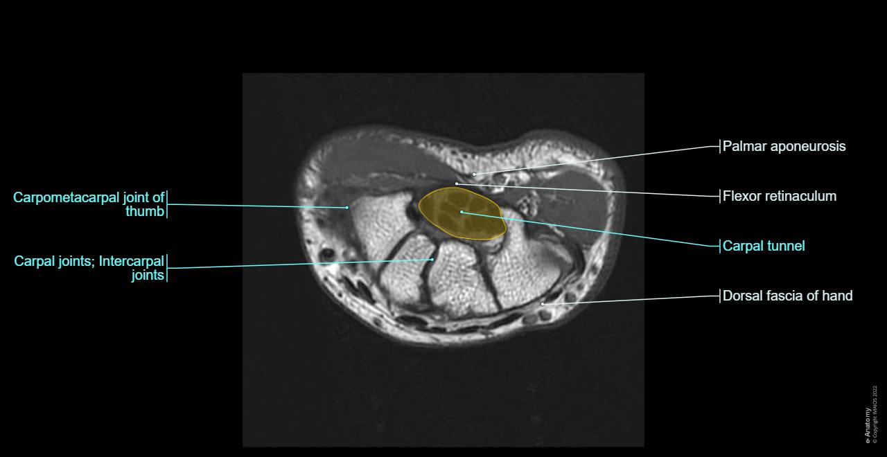
This module is a support to learn and on a daily practice of magnetic resonance imaging of the wrist, useful for a specialized audience (radiologists, surgeons, rheumatologists and physicians specializing in musculoskeletal imaging).
Images of the wrist on 3T MRI and 3D
The MRI images of the wrist are from a healthy volunteer. Image acquisition was performed on a 3 Tesla MRI with a thickness of 4 mm in contiguous sections. We used the commonly used weightings (T1 and RFP-FatSat) and impacts (axial, coronal and sagittal) in medical imaging studies of the wrist.

The 3D images of the wrist joint and carpal bones are three-dimensional reconstructions obtained from a scanner.
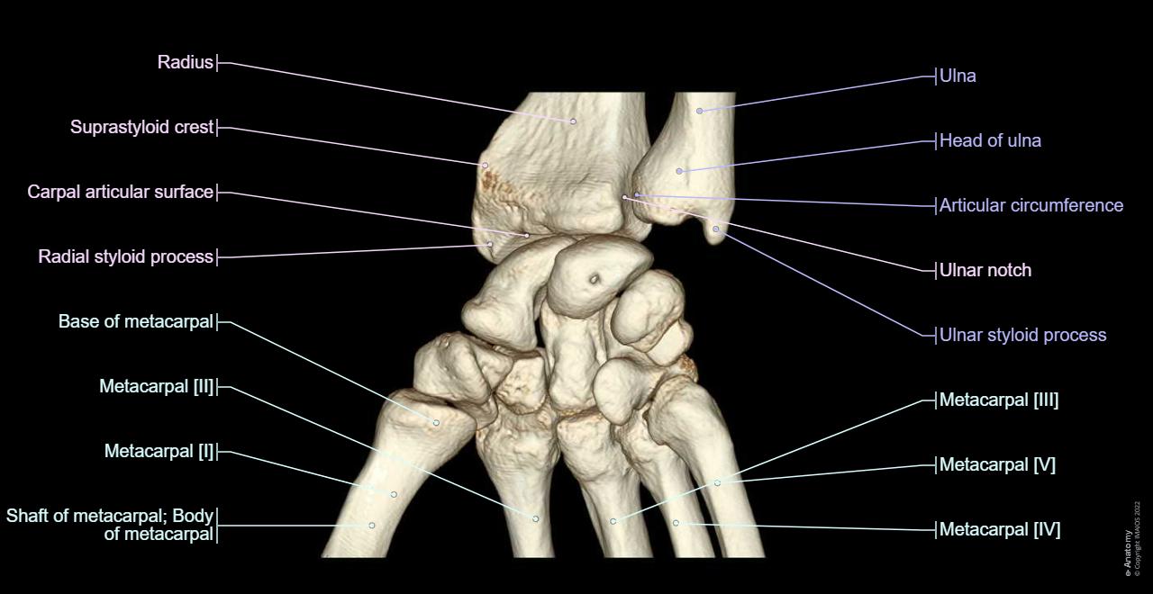
Labels for the anatomical structures of the carpus
340 anatomical structures of the wrist were labeled, accessible on "Anatomical parts":
- General anatomy: the different regions of the wrist and hand (carpal, metacarpal, hypothenar and thenar eminence).
- Bone: the anatomy of the bones of the wrist, subdivided into radius, ulna, carpal bones (scaphoid, lunate, pisiform, triquetrum, hamatum, capitatum, trapezoid and trapezium) and the metacarpal bones.
- Articulation: the different joints of the carpus, including the distal radioulnar joint, the radiocarpal, midcarpal and carpometacarpal articulations.
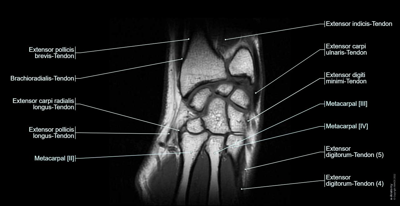
- Ligaments: all the ligaments of the wrist visible on an MRI are shown on this anatomy module, including collateral ligaments, the radiocarpal and ulnocarpal ligaments as well as the intercarpal ligaments.
- Muscles: mainly the muscles of the forearm and the palmar region (muscles of the little finger, thumb muscles).
- Tendons: the many tendons of the wrist are all labeled on this picture, from the tendon of the flexor carpi radialis to the flexor digitorum profundus tendon.
- Fascia / aponeuroses: notably displays the transverse carpal ligament (flexor retinaculum) and the palmar fascia. The carpal tunnel and ulnar canal (Guyon's) are visible by selecting the different filters on "Anatomical parts".
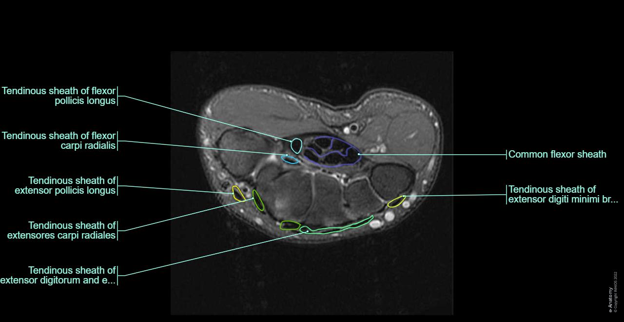
- Carpal tendon sheaths: describes the various synovial sheaths surrounding the tendons of the wrist: sheath of the extensor muscles of the fingers, sheath of the flexor carpi radialis, tendon sheath of the long thumb flexor...
- Arteries: the radial and ulnar arteries are detailed on this MRI of the wrist, as well as their branches and the various dorsal and palmar arches.
- Veins: represented by the superficial venous system of the hand (dorsal hand veins, basilic vein, cephalic vein) and the deep venous system of the wrist (branches of the ulnar and radial veins).
- Nerves: shows the innervation of the wrist by its main nerves and their branches (ulnar, median and radial nerves).
Anatomical parts
On "Anatomical parts" the user can choose to hide or show all the anatomical labels of the wrist region or to display only the most important anatomical structures in order to avoid the overloading of the images.
Contrast
The user can choose T1 or DP-FatSat weightings on this MRI scan of the wrist.
Anatomical terminology
The anatomical structures were labeled by Antoine Micheau M.D. (Radiologist, Montpellier – France), using the Terminologia Anatomica.
No content available
