

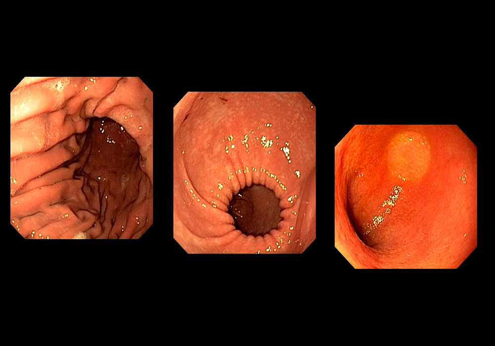
Normal gastroduodenoscopy exam of a dog
Normal gastroduodenoscopy exam of a dog
Introduction
An endoscopy allows direct observation of the gastrointestinal tract lumen, thorough and non-invasive evaluation of potential lesions, specific diagnosis through mucosal biopsy and potentially the removal of foreign bodies.
Gastroscopy and duodenoscopy are the most common procedures performed in small animals by veterinary gastroenterologists. The indications are numerous: regurgitation, vomiting, haematemesis, dysphagia, etc.
Material and methods
This gastroduodenoscopy was performed on a dog by Juan Hernandez, DVM, Dipl. ACVIM and ECVIM-CA, Head of the Department of Internal Medicine, Oniris University Veterinary Hospital, Nantes - France. The abdominal esophagus, stomach (body, cardia, fundus, pyloric antrum, angular incisure) and the first part of the duodenum were explored.
The anatomical structures were labeled by Stephan Mahler, DVM, MSc, PhD (Veterinary Anatomist - IMAIOS), according to the Nomina Anatomica Veterinaria (NAV).
They have been grouped into three different sections:
Esophagus
Stomach
Duodenum
No macroscopic abnormalities were observed during this examination.
No content available
International Committee on Veterinary Gross Anatomical Nomenclature. 2017. Nomina Anatomica Veterinaria. Sixth Edition. Ghent (Belgium).
Evans HE, de Lahunta A. 2013. Evans Miller’s Anatomy of the Dog. Fourth Edition. St Louis (MI): Elsevier Saunders.
Tams TR, Rawlings CA. 2010. Small Animal Endoscopy. Third Edition. Elsevier Mosby.
