

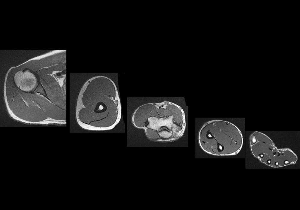
MRI of the upper limb anatomy - Atlas of the human body using cross-sectional imaging
MRI of the upper limb anatomy - Atlas of the human body using cross-sectional imaging
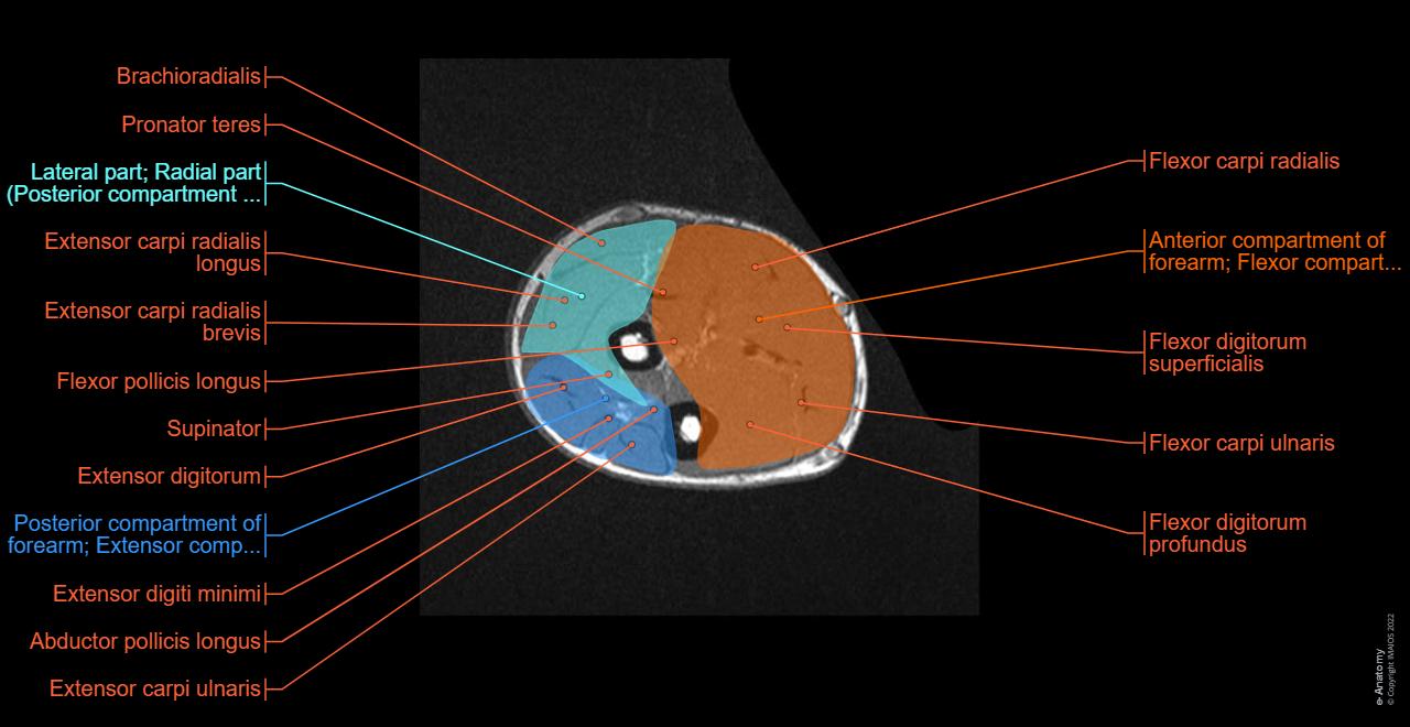
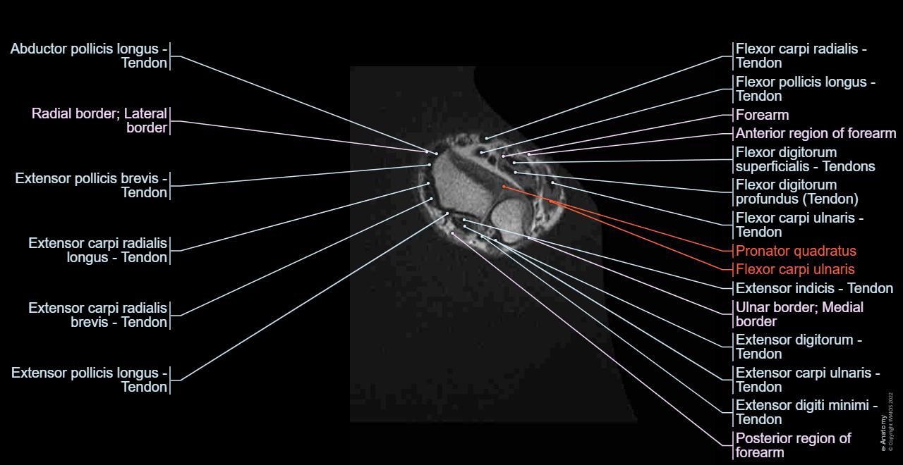
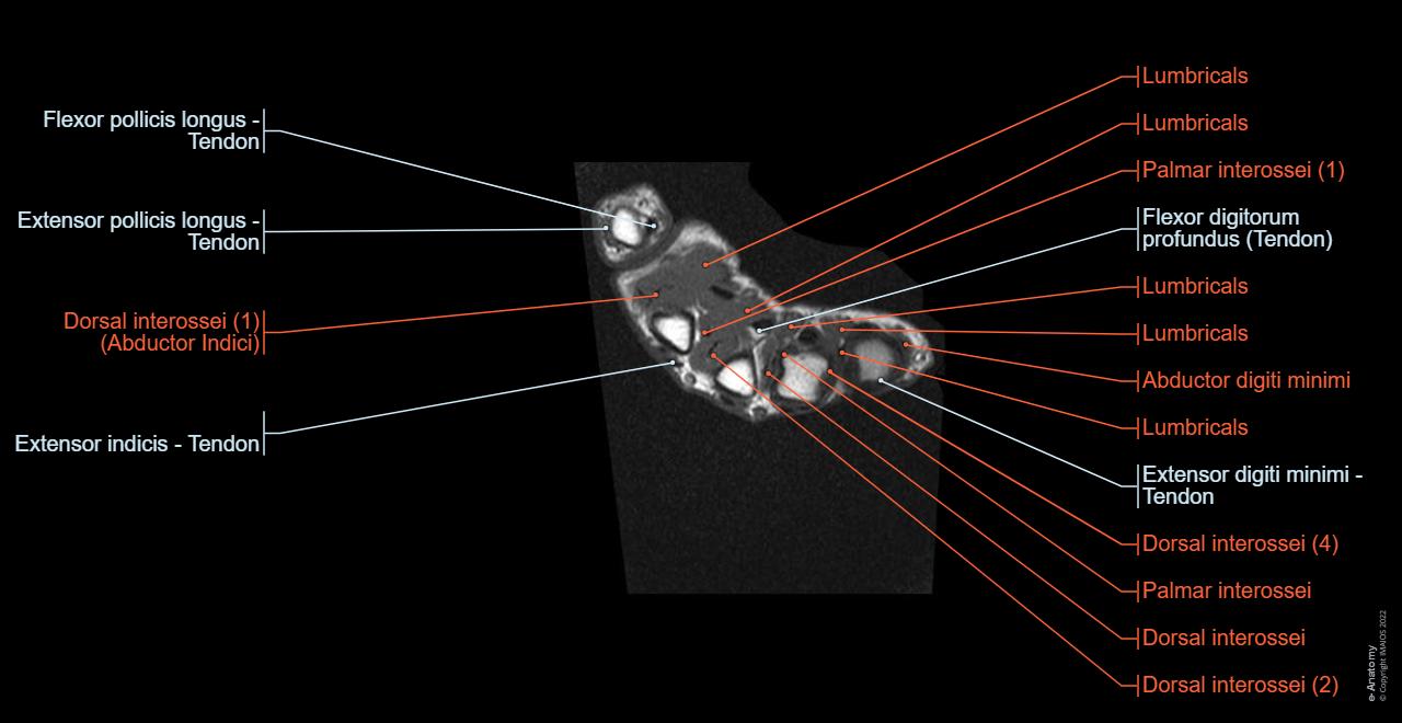
We created an anatomical atlas of the upper limb, an interactive tool for studying the conventional anatomy of the shoulder, arm, forearm, wrist and hand based on an axial magnetic resonance of the entire upper limb. Anatomical structures and specific regions are visible as dynamic labeled images.
Cross-sectional anatomy: MRI of the upper limb
An MRI was performed on a healthy subject with a spin-echo T1 weighted acquisition.
Data and DICOM images (archived on a PACS (Picture Archiving and Communicating System) were processed and exported as JPEG images. Adobe Animate and Adobe Photoshop allowed us to develop an atlas-based application with useful features and user-friendly interface to explore the skeleton anatomy.
Anatomical structures were labeled using the Terminologia Anatomica.
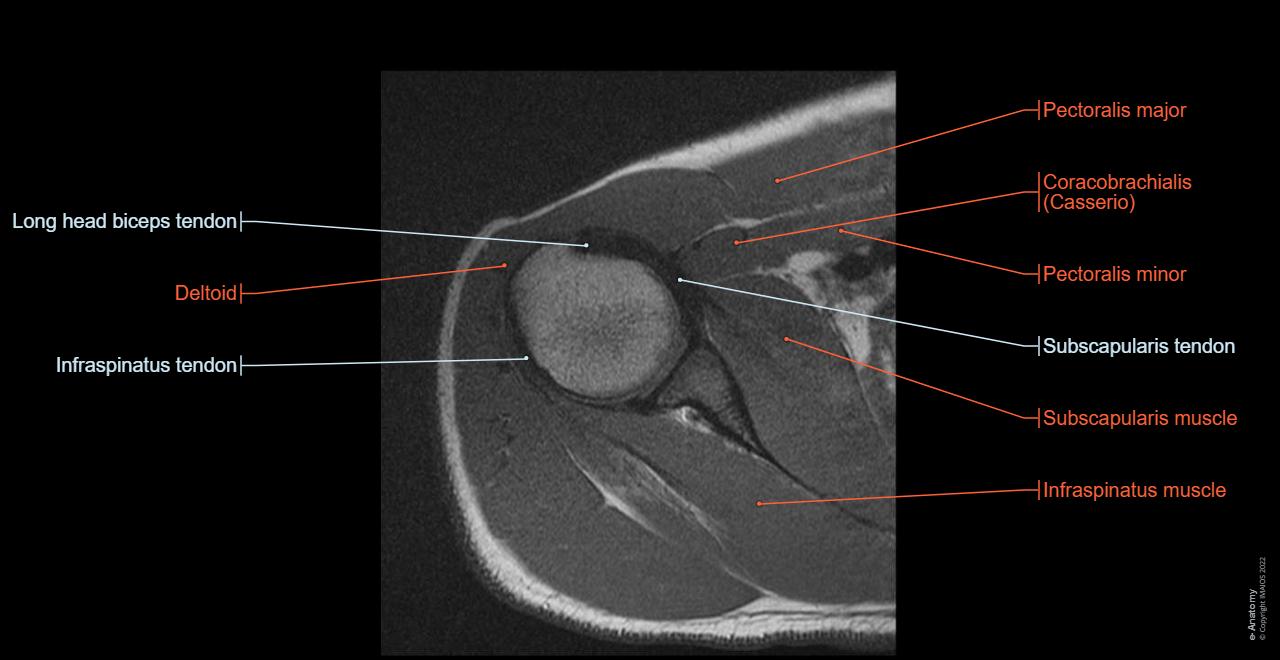
Anatomy of the arm, forearm, wrist, shoulder and hand: how to view the anatomical labels
This module is a comprehensive and affordable learning tool for medical students and residents and especially for rheumatologists, orthopedic surgeons and radiologists.
An atlas containing images on the axial planes, allowing the user to learn and review orthopedic anatomy interactively. The images were labeled, providing an invaluable teaching resource. The quiz mode allows the evaluation of user progress.
On "Anatomical parts" the user can choose between the different groups of anatomical structures:
- Arteries
- Veins
- Muscles
- Tendons
- Bones
- Nerves
- Ligaments
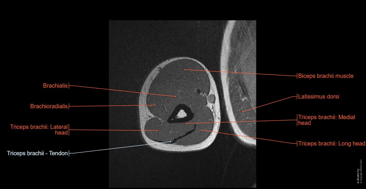
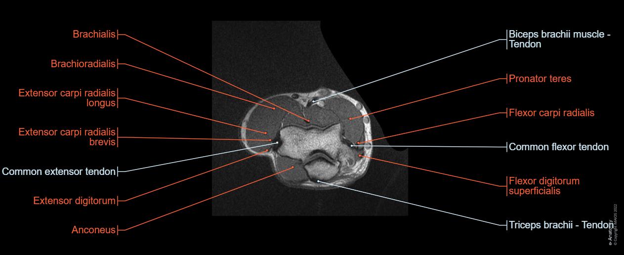
By moving the mouse cursor over a particular area of the arm or forearm, this area is highlighted and the labels are displayed: anterior, lateral or posterior compartment.
On the vertical left menu, a medical illustration of an upper limb skeleton based on a three dimensional (3D) model simplifies the access to the anatomical regions.
No content available
