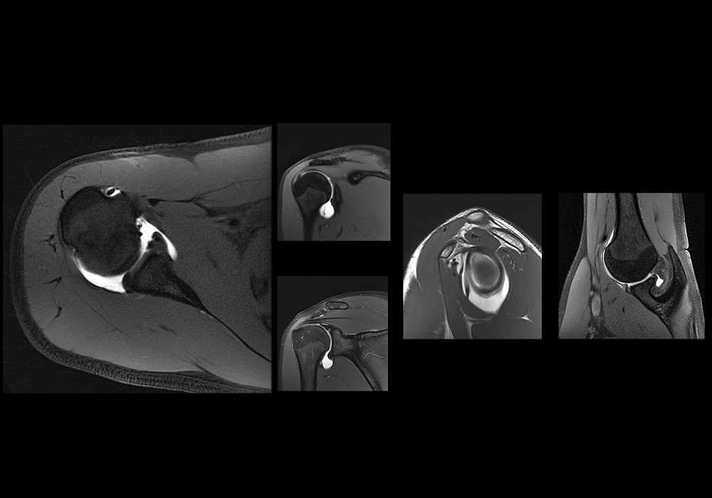
肩部磁共振关节造影:盂肱关节和肩袖解剖磁共振
前言
本文件包含关于订阅付费服务条款(以下简称“条款”)的重要信息,这些服务可通过网站www.imaios.cn(以下简称“网站”)或由医脉欧(上海)信息咨询有限公司(以下简称“IMAIOS”或“发布方”或“许可方”)发布并在Apple Store和安卓平台上提供的应用程序(以下简称“应用程序”)获得。在此情况下,由于应用程序可通过这些平台获取,使用应用程序的客户亦需接受Apple Store或Android平台的使用条款。本条款仅适用于根据法律和判例定义的消费者,且其行为完全出于个人目的。根据中华人民共和国(以下简称“中国”)法律,付费服务的基本特征和价格可在网站和应用程序中查阅。
本条款可在下单订阅前及完成订单前通过网站和应用程序获取。
发布方信息
公司名称:医脉欧(上海)信息咨询有限公司
公司形式:有限责任公司
注册地址:上海市静安区南京西路1266 号2幢1578室,邮编:200040
统一社会信用代码:91310000MAE3FRX513
法定代表人:Nguyen-Thanh Denis HOA
电子邮件:contact@imaios.cn
1. 合同的目的
1.1 定义
为本合同之目的,以下术语定义如下:
“许可”指本许可协议,该协议定义了发布方规定的访问和使用产品的条款,客户需接受这些条款以提交订单。根据本许可的条款,发布方向客户授予使用软件的许可,仅供客户为自用目的和自身需求使用。本术语指本合同及其附件和后续修改。
“被许可方”或“客户”或“用户”:指希望根据本许可规定的条款访问和使用IMAIOS提供的付费服务的自然人,且以其个人名义行事。
“软件”或“解决方案”:指由IMAIOS发布和分发的原创软件(包括但不限于e-Anatomy和vet-Anatomy),被许可方希望访问其付费功能。该原创软件基于无形元素和组件,例如源代码的开发、一组指令、程序(如图形界面、数据库等)、流程和规则,这些元素和组件是特定性和创造性选择的结果,尤其是智力贡献的结果。本术语还包括与所述产品及其附件(如图像、文本等)相关的文档,即任何附着于软件或与软件相关的、使其运行和得以使用的元素。
“付费服务”:指通过网站或应用程序提供的对软件功能的付费访问。许可方在此授予被许可方一项被其接受的在整个许可期限内非独占、不可转让、不可分配、可撤销的许可,以使用所订购的付费软件功能,且仅根据其预期目的和个人需求使用。
1.2 目的
本合同的目的是确定客户的订阅条款,使其能够在许可规定的使用范围内通过网站或应用程序访问产品的付费服务。
1.3 范围
被许可方声明其已阅读了IMAIOS网站上可自由访问的文档,该文档介绍了软件的特征以及发布方授予客户使用软件的许可条款和条款。若订单通过Apple Store或安卓平台提交,被许可方亦声明其已阅读了相关使用条款。
被许可方确认,授予用户的使用权限仅允许用户通过网站或下载到其移动设备(如平板电脑、手机)上的应用程序查阅软件。被许可方同意,本许可不包括除第1.1条“定义”中所述的“付费服务”之外的任何附加服务。本许可未授权被许可方另行授予分许可。
本许可不包含对被许可方的用户支持。
被许可方在此同意,软件包含受适用知识产权及其他法律(包括但不限于版权)保护的专有内容、信息和材料,被许可方不得以任何方式使用此类专有内容、信息或材料,除非根据本协议条款属于受许可的使用。
许可方在商标、设计和模型方面的知识产权不属于本许可的范围。
2. 订购条款
为了订阅付费服务,客户需遵循以下步骤:
2.1. 使用网站访问时
a. 客户需点击链接或输入网址
b. 客户需遵循网站的指示,特别是与客户注册相关的指示,若客户尚未拥有用户账户,则需创建账户:在此情况下,客户需提供与其职务、姓名、姓氏、账单地址、电子邮件地址和电话号码相关的信息。客户同意提供真实且准确的信息。客户可随时通过访问其账户修改其个人信息、登录名和密码。客户对其登录名和密码的使用负全部责任,并同意对其保密。若账户丢失或未经授权使用,客户需立即通过网站上所有页面上注明的“联系我们”功能通知发布方。若客户有可能丢失密码,可在登录输入用户名和密码时点击链接,要求发布方为其生成一个新密码,该密码将发送至其个人信息中指定的电子邮件地址。
c. 此后,客户需阅读《订阅条款与条件》链接中的内容以及网站和应用程序的访问与使用条款。接下来,客户需勾选“我接受《订阅条款与条件》及《访问与使用条款》”选项。
d. 希望访问付费服务的客户需填写订单表。若在连接期间长时间无操作,客户在此无操作之前选择的付费服务可能无法保留。客户需重新开始选择付费服务。客户需核对订单内容,并在必要时识别并纠正错误;在确认订单之前,客户可随时放弃订单。
e. 客户需确认订单并遵循支付服务提供商的指示支付总价。
f. 客户将被重定向至“我的订单”页面。同时,客户将收到一份电子订单确认函,其中显示订单编号和支付价格。客户可通过其账户访问电子格式的发票。我们建议用户打印订单确认页面,以保留订单内容及订单编号。此编号需妥善保存,若后续索赔将会用到该编号。
2.2 使用应用程序访问时
a. 客户需前往应用程序,或若尚未拥有应用程序,则需前往Apple Store或Android平台下载应用程序,并同意遵守相关平台的条款和条款,以及网站和应用程序的访问与使用条款。
b. 客户需在应用程序内创建用户账户,以便在其他设备如网站或其他平台上的同一应用程序上享受付费服务。为此,客户需提供其职务、姓名、姓氏、账单地址、电子邮件地址和电话号码等信息。客户同意提供真实且准确的信息。客户可随时通过访问其账户修改其个人信息、登录名和密码。客户对其登录名和密码的使用负全部责任,并同意对其保密。若账户丢失或未经授权使用,客户有责任立即通过应用程序内的“联系我们”选项卡通知发布方。若客户丢失密码,可在登录输入用户名和密码时点击链接,要求发布方为其生成一个新密码,该密码将发送至其个人信息中指定的电子邮件地址。
c. 无论何种情况,付费服务的订阅均通过平台完成。因此,除了阅读“订阅条款”链接中的内容以及网站和应用程序的访问与使用条款,并勾选“我接受《订阅条款与条件》及《访问与使用条款》”外,客户还需遵守Apple Store、安卓平台或支付服务提供商的通用条款和条件,这些条款和条件已在客户下载应用程序时已阅读并需再次接受。
d. 接下来,希望访问付费服务的客户可自行选择其想订阅的服务、点击“订阅”、选择所需订阅内容并点击“订阅”来订购所需服务。客户需根据平台保留的条款确认订单,并遵循平台的指示支付价格。
e. 客户将立即收到一份电子订单确认函,其中显示订单编号和支付价格。客户可通过平台内的账户访问电子格式的发票。我们建议客户保留订单确认页面,以保存订单内容及订单编号。后续任何索赔时需援引该编号。
3. 付费服务的访问与取消
3.1 访问与限制
订单经验证并付款后,客户可通过提供用户名和密码激活在网站上访问付费服务的权限。客户可通过登录网站访问已订购并支付的付费服务。应用程序上的付费服务访问权限在客户通过安卓平台、Apple Store或支付服务提供商账户在应用程序上完成支付后生效,但也可通过在应用程序内链接到网站上开设的账户,以享受在网站上订阅的付费服务。事实上,尽管客户账户的使用严格限于本人使用,不允许账户共享,但客户有权基于单一订阅,在不同设备上非同时使用付费服务。
被许可方确认,付费服务的使用不包括任何安装、适配、定制或培训服务:若需要此类服务,被许可方可向许可方提出请求,并需另行计费。
nsor.
3.2 取消
对于通过网站提交的订单,即使是法律规定之外,客户有权自完成下单之日起三十(30)天行使取消权。客户可在此期间内,无需说明任何理由,通过向发布方发送信件或电子邮件(请附上回执)明确表示取消订单。仅供参考的标准取消表格将在附录中提供给客户。退款将在十四(14)天内完成。
对于通过应用程序、安卓平台、支付服务提供商或Apple Store提交的付费服务订单,则适用相关平台的使用条款。
在此情况下,通过Apple Store和安卓平台订购服务的客户明确承认,由于合同标的的性质(客户在行使取消权之前即已请求访问和使用产品),当付费服务在Apple Store订单提交时即已下载,或通过Android平台或支付服务提供商订购的服务即时可用时,取消权将不适用。
在完成上述订单的各阶段期间,客户承诺遵守本合同的条款。
发布方保留因任何合法理由拒绝订单的权利,特别是当订单异常、出于恶意提交,或与客户就先前订单的支付存在争议时。
4. 价格与支付
4.1 价格
为访问和使用付费服务,客户需支付与所选服务及订阅期限对应的价格。
允许访问和使用付费服务的订阅价格在网站或应用程序上的当前价目表中明确规定,并在订单时再次列明;该价格已包含所有税费。
付费服务包括:
· 授予客户使用网站和应用程序的许可费用;
· 用户支持、使用统计、托管及软件带宽使用费用,如适用。
所有其他服务均不包括在价格内,包括任何可能的安装、适配、定制或培训费用:若需要此类服务,被许可方可向许可方提出请求,并需另行计费。在此情况下,与访问网站或应用程序相关的电信费用由客户自行承担。优惠和价格的有效期由网站和应用程序的更新而定。
4.2 支付
在网站和应用程序上,客户经由第三方支付处理提供商,通过信用卡或借记卡、Apple Pay、微信支付或支付宝支付总价。接受的信用卡或借记卡为第三方支付处理提供商支持的卡种。
若适用,在验证银行卡信息并收到客户使用的数字钱包或银行卡发卡公司的扣款授权后,交易将立即从客户的数字钱包或银行卡中扣除。
通过数字钱包或支付卡支付的承诺不可撤销。通过提供其数字钱包或信用卡信息,客户授权支付服务提供商从其数字钱包或信用卡中扣除与总价对应的金额。
为此,客户确认其为待扣款的数字钱包或银行卡的持有人,且银行卡上的姓名确为其本人。客户需提供信用卡的16位数字和有效期,以及如有必要情况下还需提供可视密码。
若无法扣除总价,订单将立即自动取消。
许可方力求确保在网站上传输的数据的机密性和安全性。客户知晓,当通过数字钱包或信用卡支付时,许可方使用服务提供商的服务,该服务提供商负责确保支付交易的安全性;发布方无法访问客户的信用卡号码,这些号码不存储在发布方,而是存储在支付服务提供商处。此外,当在应用程序上订阅付费服务时,发布方不处理支付操作,支付操作由平台或支付服务提供商负责。
5. 访问期限与续订
付费服务的访问权限自订单支付之日起生效,期限为客户订购时选择的期限。对于某些付费服务,个人订阅将在到期日自动续订;用户可最迟在续订前一天在其个人资料界面的“订阅”部分,或者在应用程序中Apple Store(设置 > Apple ID)、安卓平台或支付服务提供商提供的购买和/或订阅管理页面上,关闭此续订功能。
用户将通过电子邮件获知拒绝续订订阅的截止日期,该电子邮件将最早在年度订阅续订日期前3个月,最迟在续订日期前1个月发送。
双方明确同意,IMAIOS保留在不同期间修改其价格和访问条款的权利。
此外,某些付费服务可无限期订阅:IMAIOS可能不会为这些服务要求新的付费订阅,但不承诺确保这些服务或其所处的应用程序继续保持可用。
6. 适用的合同文件
通过使用IMAIOS的网站和应用程序,被许可方无条款接受IMAIOS网站上免费提供的网站和应用程序的访问与使用条款,以及许可方的隐私政策。若通用使用条款与当前合同发生冲突,以后者为准。此外,使用应用程序的客户需遵守所用平台即安卓平台、支付服务提供商或Apple Store的条款和条款。若这些条款与当前条款发生冲突,以后者为准。
7. 合同的终止
若任一方违反其合同义务,在友好协商无效的情况下,受损方可发出要求回执的挂号信要求违约方履行义务,若在30天内未得到履行,受损方可通过要求回执的挂号信通知终止合同。若被许可方未能履行其义务,将不退还已支付的费用,包括订阅剩余期限的费用。
8. 其他条款
除非有相反证明,网站“我的账户”页面上记录的数据构成所有在网站上下达的订单的证明,客户可随时访问其订单历史记录。同样,在应用程序上下达的订单可在Apple Store、安卓或支付服务提供商平台上查看。
客户可在订单时通过网站和应用程序内的链接访问双方签订的合同(订阅条款、隐私政策、网站和应用程序的访问与使用条款)。客户可下载这些合同并将其保存在其选择的其他持久介质上。
本协议对双方及其各自的承继人均具有约束力。除非获得许可方的事先书面同意,否则被许可方不得转让其在本协议或任何法律下的权利、义务或特权。
本条款的翻译版本仅供参考。若翻译版本与英文版本存在不一致,以英文版本为准,英文版本为唯一对双方具有约束力且规范与发布方关系的版本。
本合同及其解释受中华人民共和国法律管辖,双方认可发布方注册地所在上海市的人民法院具有专属管辖权。
取消表格模板
(适用于通过网站提交的订单)
致:contact@imaios.cn
医脉欧(上海)信息咨询有限公司
上海市静安区南京西路1266 号2幢1578室,邮编:200040
我在此通知贵方,我决定行使取消权,取消以下订单:
订单编号:XXXX,涉及服务:XXXX,下单日期:XX/XX/202X。
因此,我请求贵方尽快并最迟在收到本通知后14天内退还我下单时支付的金额:总计 .... 元人民币,此请求依据《中华人民共和国民法典》的规定。
消费者姓名:
消费者地址及(如适用)电子邮件地址:
日期:
请仔细阅读
1. 本文件包含了关于访问和使用网站 www.imaios.cn(以下简称“网站”)及应用程序(包括但不限于在安卓商店和Apple Store上提供的e-Anatomy和vet-Anatomy,以下简称“应用程序”)的条款和条件(以下简称“条款”)的重要信息,适用于发布网站和应用程序的公司与所有网站和应用程序用户(以下简称“用户”)之间。
2. 网站和应用程序仅可用于信息获取目的。网站和应用程序仅供具备专业知识的医疗保健专业人员及已参与独立于网站和应用程序的培训过程以成为专业人员的人士使用。
网站和应用程序无法解答公众的医疗问题。网站和应用程序并非用于替代患者与其医疗保健专业人员之间的关系,也非用于替代医疗建议。网站和应用程序未经临床使用测试或批准。网站和应用程序不是医疗设备,也未获得相关认证。网站和应用程序不得用作诊断工具。
一般而言,我们建议您在查阅任何包含医疗内容的互联网网站和应用程序之前,系统地咨询您的医生。
3. 通过访问网站或应用程序及其内容,或使用网站和应用程序中提供的任何服务,无论您是否为医疗保健专业人员,您完全且无保留地接受下文定义的这些条款,并声明您将无限期受这些条款的约束。这些条款包括各种责任限制和排除条款,以及管辖任何争议处理的司法条款。
通过访问网站或下载应用程序,您确认您已了解发布者实施的隐私与数据保护政策,该政策可通过网站任何页面及每个应用程序内的链接 https://www.imaios.cn/en/privacy-policy 访问。您声明您不反对发布者实施的此隐私与数据保护政策。
4. 这些条款可能随时更新;它们通过网站所有页面及每个应用程序内的链接系统地提醒任何访问网站或应用程序的人士。因此,我们感谢您定期查阅这些条款及其更新。
如果您不接受全部或部分条款,请您放弃使用网站和应用程序并关闭它们。
5. 这些条款仅适用于您对网站和应用程序的访问与使用;该等条款不会更改您与发布者之间可能存在的任何其他协议的条款或条件。
1 网站和应用程序的介绍
1.1 网站和应用程序的介绍与目标
网站和应用程序仅面向具备专业知识的医疗保健专业人员(以下简称“用户”)。网站和应用程序是为教育目的而设计的,并包含医疗信息(文章、插图、工具和其他资源等)
1.2 网站和应用程序的来源
网站和应用程序由医脉欧(上海)信息咨询有限公司发布,地址为上海市静安区南京西路1266 号2幢1578室,邮编:200040(以下简称“发布者”)。
发布方的董事为Denis Hoa先生。
1.3 网站的托管
网站由AWS中国(宁夏)区域托管,由宁夏西云数据科技有限公司(NWCD)运营。
1.4 定期贡献者
医脉欧团队的所有成员均定期为网站和应用程序提供医疗信息,特别是其经理:
- Denis Hoa,医学博士,毕业于蒙彼利埃医学院,放射诊断与医学影像学专业,蒙彼利埃医学院优秀毕业生,医学放射物理学与影像学硕士双研究学位持有者;与
- Antoine Micheau,医学博士,毕业于蒙彼利埃医学院,放射诊断与医学影像学专业。
1.5 网站与应用程序的资金来源
网站由发布者和用户订阅提供资金。应用程序由发布者和用户通过应用内购买提供资金,应用程序为免费增值类型。
发布者的股东与制药行业无直接或间接联系。
2 访问网站和应用程序
2.1 开放访问与注册
2.1.1 网站
网站免费且开放访问。然而,发布者保留单方面且不事先通知的情况下对网站的全部或部分访问收费的权利。
访问网站的某些区域可能需要根据说明进行注册。如有必要,发布者保留单方面且不事先通知的情况下暂停、限制或拒绝任何不遵守条款的注册用户(以下简称“注册用户”)访问网站的权利。
2.1.2 应用程序
应用程序(包括但不限e-Anatomy和vet-Anatomy)各自基于一组指令、程序和规则而创建。应用程序是发布者创建的独特源代码的表达。
这些应用程序都是由医脉欧创建的原创软件。
这些应用程序不仅仅是自动逻辑的简单实现。
每个应用程序都有编程行、代码、结构和发展语言,反映了发布者的创意选择和智力贡献。
应用程序可免费在安卓、Apple Store或其他应用平台上下载。然而,某些选项并非免费,发布者保留单方面且不通知的情况下对最初免费的功能收费的权利。
2.2 网站和应用程序及其内容的更新、中断与可用性
发布者可能随时修改或删除网站和/或任何应用程序上提供的信息。发布者保留为维护或其他原因暂时或永久中断或修改对网站和应用程序的全部或部分访问的权利,且此类中断不会引发任何义务或赔偿。对应用程序的访问也可能因安卓或Apple Store的单方面决定而中断或暂停。
产品和服务规格可能随时更改且不另行通知。此外,医脉欧不保证在线或应用程序上列出的产品或服务在您订购时可用。
2.3 范围
在此明确同意,授予的访问权限仅允许用户访问网站和应用程序。
用户承认这些条款未授予用户对网站和应用程序的任何知识产权(特别是商标、设计或模型)的所有权。
3 用户的特别承诺
通过使用网站和应用程序,用户特别同意其不得:
- 与其他用户共享其资质证明,每个账户仅供严格个人使用;
- 干扰或破坏网站和应用程序及其内容、资源(与网站连接或通过网站访问的服务器或网络)的安全性;
- 干扰或破坏其他用户的体验
- 通过或向任何网站上上传、发布或以其他方式传输任何病毒或其他有害文件;
- 通过网站或应用程序向未同意接收邮件的人或实体发送任何类型的未经请求的群发邮件;
- 试图未经授权访问网站或应用程序或其受限部分;
- 使用网站或应用程序寻求、提供或获取特定医疗建议、医疗意见或诊断;
- 使用网站或应用程序寻求、提供或获取与特定健康相关检查相关的具体答案和/或课程。
4 提供信息的质量与使用
4.1 质量
发布者尽力确保网站和应用程序上提供的信息质量及其定期更新。然而,网站和应用程序可能包含错误或不准确的信息、遗漏或非基于发布者意愿发布的数据。
发布者不承担更新网站和应用程序上的信息的义务。
网站和应用程序上发布的数据明确提及其来源,并在必要时提供该来源的超链接。最后修改日期清楚地显示在网站或应用程序的相关页面上。
4.2 使用
发布者特别提醒,网站和应用程序上发布的信息仅具指示性和教育目的,不提供任何明示或默示的保证,仅供在法国、中国或其原籍国获得执业资格的医学专业人员及医学学生使用,但某些应用程序可能面向更广泛的公众。
此信息绝不替代医学专业人员的意见,也不得被视为或解释为任何类型的建议或推荐。
用户不得在任何情况下使用网站或应用程序描述病情、做出诊断、决定治疗或做出任何与患者治疗相关的医疗决策。
用户在任何情况下均对其使用其可获取的信息全权负责。我们建议用户谨慎使用该信息,并运用其专业判断进行评估。发布者还建议系统查阅信息的其他来源。
5 产品或服务的可用性
网站或应用程序上任何援引的发布者的产品、应用程序或服务之处,并不意味着该产品或服务在您所在国家/地区可用,且可能受到不同法规和使用条款的约束。该等援引并不意味着发布者有意在用户所在国家/地区销售此类产品、应用程序或服务。网站和应用程序包含有关在全球范围内可能或不可用的产品和服务的信息。用户在使用网站、应用程序或通过网站和应用程序访问的产品/服务之前,必须确保此类使用不违反其居住国的法律。
用户全权负责验证网站、应用程序(或通过网站或应用程序访问的产品/服务)的内容是否符合其访问网站或应用程序所在国家/地区的法律。因此,用户不得以违反其访问网站或应用程序所在国家/地区法律、法规或道德规定的方式使用网站或应用程序。发布者或任何其他参与网站或应用程序创建和运营的第三方均不对因不遵守使用网站或应用程序所在国家/地区法律而承担责任。
6 责任与保证
6.1 发布者责任与保证的排除
发布者及其合作伙伴、员工或任何其他参与网站和应用程序创建和运营的第三方均不对用户访问、使用或解释网站或应用程序上的信息所导致的任何直接或间接损害或损失承担责任。
6.2 用户的责任与保证
用户保证并赔偿发布者及其合作伙伴、员工或任何其他参与网站或应用程序创建和运营的第三方,因用户使用网站或应用程序或与其使用直接或间接相关的任何有害后果而导致的任何第三方行动或索赔。
用户因此承担发布者及其合作伙伴、员工或任何其他参与网站和应用程序创建和运营的第三方可能被判决的全部损害赔偿,以及相关的法律费用和所涉及的费用。
6.3 第三方网站的超链接
网站和应用程序可能包含指向由第三方管理的其他互联网网站的超链接。然而,发布者无法定期验证这些链接网站的质量。发布者不对这些网站的内容或这些网站上提供的服务承担责任。
此外,对第三方网站的摘要和/或链接并不意味着发布者认可该网站或其上的产品或服务。发布者不保证此类第三方网站上引用的任何内容的准确性,且不对访问或无法访问这些网站所导致的任何损害或伤害承担责任。
7 网站、应用程序及其内容的所有权
7.1 网站和应用程序内容的保护
网站、应用程序及其内容(以下简称“内容”)的所有知识产权,包括文本、数据库、软件、应用程序、幻灯片、徽标、图像、绘图、图形、动画序列、声音、视频,均归发布者或已授权发布者使用该内容的第三方所有。
该内容整体受法国和国际法律保护,特别是有关版权、设计与模型、商标和数据库的法律。
网站和应用程序上提及的名称和品牌为发布者或其受益人的注册商标。
用户在此明确同意,发布者授予的访问权限不得解释为向用户授予发布者持有的知识产权的许可。因此,任何复制、模仿及更广泛地对这些商标的利用均受禁止。
7.2 网站和应用程序的使用——授予的许可
网站和应用程序仅供用户个人自用。
发布者授予用户全球范围内、非独占、不可转让、不可转移、可撤销的许可,以在本协议期限内及规定的条件下使用网站和应用程序。用户承认网站和应用程序包含受适用知识产权法律保护(特别是版权法)的独家内容、信息和材料。IMAIOS持有网站和应用程序的知识产权,包括版权。用户仅获得使用权,不涉及所有权转让。
用户在同时符合以下条件的情况下可打印或下载网站页面,或打印或截取应用程序页面:
- 部分且合理地打印、下载或截取(即网站或应用程序的10%以内);
- 不删除内容中的任何所有权声明,不修改内容;
- 仅将打印、截取或下载的信息用于用户的个人自用用途,不用于商业目的;
- 不得公开使用或分发打印、截取或下载的内容。
任何其他用途(特别是任何为商业目的复制、展示、修改、改编、分发,无论是否营利)均严格禁止,除非事先获得发布者的书面同意。
发布者作为网站和应用程序上现有或使用的全部或部分数据库的生产者和所有者,严格禁止提取和使用网站和应用程序上出现的数据库的全部或部分内容。
7.3 网站的超文本链接
未经发布者事先授权,不得设置指向网站内文档或页面的直接链接。
所有指向网站的链接必须经IMAIOS书面批准,除非符合以下情况:
- 链接为指向网站主页的文本链接,而非网站其他页面;
- 链接必须全屏显示网站主页,而非局部框架;
- 链接的外观、位置及其他方面不得造成与发布者或其活动或产品相关的错误印象。
无论如何,发布者保留随时无理由或无通知撤回其对链接的同意。
8 其他条款
8.1 期限
发布者可随时终止、修改、暂停或中断对网站或应用程序的任何访问和使用。发布者可删除或修改网站或应用程序的任何内容,并可限制某些功能和服务或限制对网站或应用程序的全部或部分访问,且不承担任何通知或责任。发布者保留随时单方面终止使用网站授权的权利。
8.2 投诉
所有对于随意使用(例如关于网站上有争议的贡献)或知识产权侵权的投诉和举报必须以书面形式发送至下文所述的联系地址。
如涉及知识产权侵权,必须提供以下信息:
- 声称权利受侵犯的权利人的身份、联系方式和签名;
- 权利人的授权代表的授权书(如有);
- 详细描述不尊重权利人权利并要求从网站或应用程序中删除的内容;
- 声明确认提供给发布者的信息的准确性。
8.3 用户违约
发布者未对用户对任何法律义务或本条款的违约行为采取行动的事实,不得被解释为未来放弃该义务或日后对该违约行为采取行动的权利。
如本条款的任何规定因任何法律或其他法律规则无效,则该规定视为未写入,但不会使本条款整体无效。
8.4 适用法律与管辖权
网站和应用程序设计于法国,托管于中华人民共和国。本条款受中华人民共和国法律管辖。如因本条款的应用或解释或更广泛地因任何个人或法人实体使用网站和应用程序而产生争议,各方明确同意发布者注册地上海静安区法院具有唯一管辖权,即使存在多个被告或第三方索赔。
8.5 联系方式
医脉欧(上海)信息咨询有限公司
地址:上海市静安区南京西路1266 号2幢1578室,邮编:200040
电话:+33 9 72 10 11 10
邮箱:contact@imaios.cn
个人信息保护政策
Personal Information Protection Policy
本个人信息保护政策(本“政策”)于2025年6月20日更新并生效。
This personal information protection policy is updated and takes effect on June 20, 2025.
医脉欧(上海)信息咨询有限公司(定义见下文第1条,以下简称为“我们”或“IMAIOS”)深知个人信息对您的重要性,并会尽全力保护您的个人信息安全可靠。本政策介绍了我们如何收集、存储、使用、传输、对外提供您的个人信息以及您对您的个人信息所享有的各项权利及其实现路径。
IMAIOS (Shanghai) Information Consulting Co., Ltd. (see the definition in Article 1 below, hereinafter referred to as “We” or “IMAIOS”) know the importance of personal information to you and will try our best to ensure security of your personal information. This privacy policy introduces how we collect, store, use, transmit and provide your personal information externally as well as your rights to the personal information and the path to its realization.
本政策适用于医脉欧(上海)信息咨询有限公司的产品和服务,包括但不限于https://www.imaios.cn(以下简称“网站”)及应用程序e-Anatomy和vet-Anatomy(可在 Android、Apple Store 及其他平台上使用,以下简称“应用程序”)。本政策可能根据相关法律法规和IMAIOS的业务调整不时更新。因此,我们欢迎您关注我们不时公布的最新版本的政策,随时了解可能的更新。如果您对本政策有任何疑问、意见或建议,可根据本政策“联系我们”部分所列的联系方式与我们联络。
This Policy applies to the products and services of IMAIOS (Shanghai) Information Consulting Co., Ltd., including but not limited to, https://www.imaios.cn (the “Website”) and the mobile apps e-Anatomy and vet-Anatomy (available on Android, Apple Store and other platforms), Apple Store and other platforms, hereinafter referred to as “Apps”). This Policy may be updated from time to time in accordance with relevant laws and regulations and IMAIOS's business adjustments. Therefore, we welcome you to check the latest version of the Policy posted from time to time for possible updates. If you have any questions, comments or suggestions regarding this Policy, you may contact us at the contact information listed in the “Contact Us” section of this Policy.
1. 定义
Definition
“用户”:指同意本网站和应用程序的访问和使用条款的任何自然人或法人实体。
“User”: Any natural person or legal entity who agrees to the terms of access and use of the Website and the Application.
“服务”:由IMAIOS提供和操作的工具和/或平台和/或软件,包括任何IMAIOS通过网站、应用程序等向您提供的服务。
“Services”: the tools and/or platforms and/or software provided and operated by IMAIOS, including any IMAIOS services offered to you through the Website, APPs, etc. provided by IMAIOS to you.
“网站”: https://www.imaios.cn。
“Website”: https://www.imaios.cn.
应用程序:IMAIOS发布的e-Anatomy和vet-Anatomy等应用程序。
APP: Mobile applications such as e-Anatomy and vet-Anatomy published by IMAIOS.
“关联公司”:受控于IMAIOS、控制IMAIOS或与IMAIOS共同受同一实体控制的任何公司。控制指的是持有至少50%的股权或投票权。
“Associated company”: Any company that is controlled by, controls, or is under common control with IMAIOS by the same entity. Control is defined as holding at least 50% of the equity or voting rights.
“访客”:任何访问网站和应用程序的个人。
“Visitor”: Any individual who accesses the website and APP.
“个人信息”:以电子或者其他方式记录的与已识别或者可识别的自然人有关的各种信息,不包括匿名化处理后的信息。
“Personal information”: Various information related to identified or identifiable natural persons, recorded electronically or otherwise, other than anonymized information.
“个人信息处理者”:指在个人信息处理活动中自主决定处理目的、处理方式的组织。
“Personal information processor”: Refers to an organization that autonomously determines the purpose and manner of processing in personal information processing activities.
“匿名化处理”:指通过对个人信息的技术处理,使得个人无法被识别,且处理后的信息不能被复原的过程。个人信息经匿名化处理后所得的信息不属于个人信息。
“Anonymized processing”: Refer to the process in which personal information is processed technically to make individuals impossible to identify and the processed information can’t be recovered. The information obtained from anonymized processing doesn’t belong to personal information.
2. 本个人信息保护政策的适用范围
Scope of Application of this Personal Information Protection Policy
本政策适用于网站和/或应用程序以及您可能与IMAIOS进行的其他互动(例如,在线提交表单或电子邮件往来等)。
This policy applies to the website and/or APPs and other interactions you may have with IMAIOS (e.g., online form submissions or email exchanges, etc.).
请在使用我们的网站和/或应用程序或与我们进行其他互动前,仔细阅读并了解本政策,特别是以粗体标识的条款。如您选择使用或继续使用我们的服务和/或网站或与我们进行其他互动,即意味着您完全理解本政策的全部内容,并同意我们按照本政策处理您的相关信息。如果您不同意本政策的内容,请勿使用网站和/或应用程序,并停止与IMAIOS的任何互动。
Please read and understand this Policy, especially the terms identified in bold, carefully before using our websites and/or APPs or otherwise interacting with us. By choosing to use or continue to use our Services and/or Website or otherwise interact with us, you are implying that you fully understand this Policy in its entirety and that you consent to our processing of information about you in accordance with this Policy. If you do not agree with the contents of this Policy, please do not use the Website and/or the APP and cease any interaction with IMAIOS.
本政策不适用于您通过网站和/或应用程序或者与我们的其他互动使用第三方提供的服务,请您在使用所有第三方的服务前了解相关第三方的政策,我们在服务、网站或者与您的其他互动中使用第三方的服务,并不表示我们认同第三方的政策或为第三方的个人信息处理活动提供任何保证。
This Policy does not apply to your use of services provided by third parties through the Websites and/or APPs or other interactions with us, please be aware of the relevant third party's policies before using all third party's services, and our use of a third party's services in connection with the Services, the Websites, or in other interactions with you does not imply that we subscribe to the third party's policies or provide any warranty for the third party's personal information processing activities.
3. 我们如何收集和使用您的个人信息
How we collect and use your personal information
我们的网站和应用程序免费开放并可自由访问和下载,通常无需创建客户账户,但某些服务的实现除外。作为个人信息处理者,我们通过以下方式收集个人信息:
Our website and APPs are free, open, and freely accessible, and downloadable, and do not typically require the creation of a customer account, except to enable certain services. As a personal information processor, we collect personal information through the following means:
● 当您通过创建或更新帐户、订阅服务、填写表格或通过支持或反馈渠道与我们联系而自愿提供信息时;
When you voluntarily provide information by creating or updating an account, subscribing to services, completing forms, or contacting us through support or feedback channels;
● 当您使用我们服务的过程中需要身份验证或个性化的特定功能时(访问高级资源);
When you use specific features of our services that require authentication or personalization (accessing premium resources);
● 通过自动化手段(包括cookies和类似的跟踪技术)收集,以帮助我们改进服务功能,分析使用模式,增强用户体验;
Through automated means, including cookies and similar tracking technologies, which help us improve service functionality, analyze usage patterns, and enhance user experience;
● 当您启用并授权集成时,通过您连接的第三方服务或平台收集。
From third-party services or platforms you connect with, when such integration is enabled and authorized by you.
这些个人信息是根据适用的法律收集的,并且仅出于本政策中明确声明的目的。
This personal information is collected in accordance with applicable laws and solely for the purposes explicitly stated in this Policy.
(1)由IMAIOS的用户主动提供的个人信息
Personal information voluntarily provided by IMAIOS users
当您订阅或使用IMAIOS服务时(包括但不限于:创建用户账户以访问服务;IMAIOS为用户提供与服务相关的支持时);或者当您订阅时讯简报、参与IMAIOS组织的各种活动(包括营销活动、讨论组、比赛、满意度调查、网络研讨会等)或与IMAIOS的社交网络账户进行互动或以其他方式与IMAIOS交流时;或者当您通过网站、应用程序等渠道上公布的联系方式,以电子邮件或任何其他方式联系我们,尤其是为了获取有关IMAIOS产品或服务的信息时,我们将收集您提供的姓名、电话号码、电子邮箱、地址、职业、机构或公司,您理解此类信息是为向您提供相应的资讯信息或服务所必需。如您拒绝提供您的上述信息,我们可能无法向您提供您需要的相关资讯信息或服务并与您有效互动,但不影响您使用IMAIOS向您提供的其他服务或与IMAIOS进行其他互动。
When you subscribe to or use the IMAIOS Services (including, without limitation, when you create a user account to access the Services; when IMAIOS provides support to its users in connection with the Services); or when you subscribe to newsletter bulletins, participate in events organized by IMAIOS (including marketing campaigns, discussion groups, contests, satisfaction surveys, webinars, etc.) or interact or otherwise communicate with IMAIOS' social network accounts; or when you contact us by email or any other means through the contact information published on the Website and other channels, in particular in order to obtain information about IMAIOS' products or services. when you interact or otherwise communicate with IMAIOS; or when you contact us by e-mail or any other means through the contact information published on the website, application, etc., in particular in order to obtain information about IMAIOS' products or services, we collect the name, telephone number, e-mail address, address, occupation, organization or company that you provide, and you understand that such information is used in order to provide you with You understand that such information is necessary to provide you with appropriate information or services. If you refuse to provide your information as described above, we may not be able to provide you with the relevant information or services that you need to interact effectively with us, but this will not affect your ability to use other services offered to you by IMAIOS or to interact with IMAIOS.
(2)在您使用网站及/或服务过程中记录的个人信息
Personal information recorded in the course of your use of the Website and/or the Services
当您访问我们的网站和使用我们的服务时,为了维护我们网站、服务的正常运行,改进及优化我们的网站、服务体验以及保障网络操作环境安全或基于遵守法律法规规定的需求,我们会自动收集一些个人信息:
When you visit our website and use our services, in order to maintain the normal operation of our website and services, improve and optimize the experience of our website and services, as well as to ensure the safety of the network operating environment or based on the need to comply with the provisions of laws and regulations, we will automatically collect some personal information.
● 元数据:元数据是指描述数据的数据,主要是描述数据属性的信息,用来支持如【指示存储位置、历史数据、资源查找、文件记录…】等功能。当用户访问服务时,系统会生成元数据以提供有关用户工作方式的额外信息,并方便访问和使用我们提供的功能。
Metadata: Metadata is the data that describes the data, mainly information that describes the attributes of the data and is used to support features such as historical data, resource lookups, file logging and so on. When a user accesses the service, the system generates metadata to provide additional information about how the user works and to facilitate access to and use of the features we provide.
● 连接和使用数据:当您访问网站和通过互联网使用服务时,IMAIOS的服务器会自动收集和存储信息。这些信息包括您的Internet协议地址(IP地址)、您在使用网站或服务之前访问的网页地址、您使用的浏览器类型及其配置和插件信息、您使用服务的日期和时间、您的语言首选项和cookie数据 (更多详细信息,请参考我们的cookie政策,见附件1)。
Connection and Usage Data: IMAIOS servers automatically collect and store information when you visit the Site and use the Services over the Internet. This information includes your Internet Protocol address (IP address), the address of the web page you visited before using the Website or the Services, the type of browser you are using and its configuration and plug-in information, the date and time you use the Services, your language preference and cookie data (for more detailed information, please refer to our cookie policy, see Appendix 1).
● 设备数据:IMAIOS将收集设备数据,包括设备类型、操作系统、设备设置、应用程序ID、唯一设备ID和停机数据,以便您下次访问网站或使用服务时更加轻松便捷。
Device Data: IMAIOS will collect device data, including device type, operating system, device settings, application ID, unique device ID, and downtime data, to make your next visit to the Site or use of the Services easier and more convenient.
● Cookie:IMAIOS将通过网站和服务上的cookie和类似技术收集数据。网站和服务中可能还包括cookie和类似的追踪技术,这些技术可能会通过第三方服务收集您的数据。如需了解更多有关IMAIOS如何使用这些技术的信息,请参考我们的cookie政策,见附件1。
Cookie: IMAIOS will collect data through cookies and similar technologies on the Sites and Services. The Sites and Services may also include cookies and similar tracking technologies that may collect data about you through third party services. For more information on how IMAIOS uses these technologies, please refer to our cookie policy, see Annex 1.
● 软件开发包(SDK)和应用程序接口 (API) 等代码:IMAIOS将通过接入由第三方提供的软件开发包(SDK)、应用程序接口 (API) 等代码或通过其他合作方式,以保障相关功能的实现与应用安全稳定的运行。请参考我们的SDK和API清单,见附件1。第三方合作方具体的数据处理情况请参见其隐私政策或相关服务说明。请注意,第三方合作方的代码可能因为其版本升级、策略调整等原因导致数据处理类型存在一定变化请以其公示的官方说明为准。
Software development kits (SDKs) and application programming interfaces (APIs) and other codes: IMAIOS will integrate software development kits (SDKS), application programming interfaces (APIs) and other codes provided by third parties or through other cooperative methods to ensure the realization of related functions and the safe and stable operation of applications. Please refer to our SDK and API list, see Annex 1. For specific data processing details of the third-party partners, please refer to their privacy policy or relevant service descriptions. Please note that the third-party partners’ code may have certain changes in data processing types due to version upgrades, policy adjustments, etc. Please refer to their official explanations.
(3)来自其他来源的个人信息
Personal information from other sources
我们也可能通过以下方式进行收集您的个人信息:
We may collect your personal information by the following ways:
第三方数据:IMAIOS可能会从其关联公司、合作伙伴或IMAIOS用于提高数据质量和相关性的其他方处接收您的个人信息,如身份识别数据或专业数据。此类数据也可以是更精确的数据,例如用于分析在线营销活动、满意度调查、招聘或电子邮件营销活动绩效的数据。我们在收集上述信息前将依法采取合理措施确保这些第三方提供给我们的个人信息符合法律的规定。
Third-party data: IMAIOS may receive your personal information such as identity data or professional data from its associated company, partners or other parties that IMAIOS uses to improve the data quality and relevance. Such data can also be more accurate data, such as the data used for analyzing online marketing campaign, satisfaction survey, recruitment or email marketing campaign performance. Before collecting above information, we will take reasonable measures to ensure the personal information provided to us by these third parties conforms to the laws.
4. 我们保存您个人信息的期限
The period for retaining your personal information
IMAIOS作为个人信息处理者,仅为实现服务目的所必需的时间存储您的个人信息(详情见下表)。当我们的服务或网站发生停止运营的情形时,我们将依照法律的规定以公告、邮件通知等形式通知您,在合理的期限内删除您的个人信息或进行匿名化处理,并立即停止收集个人信息的活动,以及关闭第三方应用服务接口,避免第三方通过服务继续收集您的个人信息。
As personal information processor, IMAIOS can only retain your personal information for the period required to achieve the service purpose (see details as below). When our Service or Website stops operation, we will notify you in the form of public announcement or email according to provisions of the law and delete your personal information or conduct anonymization within a reasonable period and stop collecting personal information and close the third-party application service interface to prevent third party from collecting your personal information through the Service.
我们网站和应用程序上收集的数据和个人信息将由我们的托管服务提供商 AWS 中国(宁夏)区域托管,由宁夏西云数据科技有限公司(NWCD)运营。
Data and personal information collected on our website and applications will be hosted by our hosting service provider, AWS China (Ningxia), and operated by Ningxia Xiyun Data Technology Co., Ltd. (NWCD).
服务目的 Purpose of Service | 期限 Term |
用于管理客户账户和其他行政任务,例如开具发票 For managing customer accounts and other administrative tasks, such as invoicing | 合同期限和法定保留期 Contract Term and Statutory Retention Period |
用于市场营销(营销活动、在线活动或贸易展会、客户满意度调查) Marketing (marketing events, online events or trade shows, customer satisfaction surveys) | 撤回同意时或注销账户时 Withdrawal of consent or cancellation of account |
进行安全调查,帮助防止安全或欺诈问题,以及潜在的滥用 Conduct security investigations to help prevent security or fraud issues, as well as potential abuse | 进行安全或欺诈调查及其解决所需的时间。如发生诉讼,可能延长 Time needed for security or fraud investigation and its resolution. This may be extended in case of litigation. |
管理通过联系表格或任何其他方式提出的请求 Manage requests via contact form or any other means | 撤回同意或处理请求 Withdrawal of consent or processing request |
网站访客的统计数据和报告 Statistics and Reports of Site Visitors | 不同保留期(请参考我们的Cookie政策) Different retention periods (please refer to our cookie policy) |
5. 我们如何向第三方共享和披露您的个人信息
How we share and disclose your personal information to third parties
在以下情况下,我们将另行通知并征求您的同意:(i) 向其他个人信息处理者提供您的个人信息,(ii) 公开您的个人信息,(iii) 处理敏感个人信息,(iv) 向境外提供个人信息,除非适用法律另有允许。我们将根据具体情况向您进行披露。我们承诺仅共享为了实现共享目的所必要的您的个人信息,且我们的共享仍受本政策的约束;如果我们要改变个人信息的使用及处理目的,我们将确保在遵守所有适用的法律依据后再进行此类变更。
We will provide further notice and seek your consent to (i) provide your Personal Information to other processors of Personal Information, (ii) disclose your Personal Information, (iii) process sensitive Personal Information, and (iv) provide Personal Information outside of the country, unless otherwise permitted by applicable law. We will make disclosures to you on a case-by-case basis. We are committed to sharing only as much of your Personal Information as is necessary to fulfill the purposes for which it was shared, and our sharing remains subject to this Policy; if we change the purposes for which the Personal Information is used and processed, we will ensure that we comply with all applicable legal authorities before making such changes.
请知悉,我们收集的数据由托管服务提供商 AWS 中国(宁夏)区域托管,由宁夏西云数据科技有限公司(NWCD)运营。
In particular, please be aware that the collected data is hosted by our hosting provider, AWS China (Ningxia) Region, operated by Ningxia West Cloud Data Technology Company Limited (NWCD).
在此背景下,我们自然遵守现行数据保护法律:我们与托管服务提供商的合作需遵守标准合同条款(您通过接受本政策确认此项内容),这些条款要求其确保高水平的数据安全,以防止未经授权的第三方欺诈性访问以及任何未经授权的数据破坏、更改或分发。
In this context, we naturally comply with current data protection laws: our cooperation with hosting service providers is subject to standard contractual clauses (which you acknowledge by accepting this policy), which require them to ensure a high level of data security to prevent fraudulent access by unauthorized third parties as well as any unauthorized destruction, alteration or distribution of data.
6. 我们如何保护您的个人信息
How we protect your personal information
IMAIOS关注您个人信息的安全。因此,IMAIOS采取了技术和组织措施来保证您个人信息的安全,并防止任何丢失或误用的风险以及任何未经授权的访问或披露。这些措施充分考量了IMAIOS收集、处理和存储的信息的敏感性以及技术水平。
IMAIOS is concerned about the security of your personal information. Therefore, IMAIOS has taken technical and organizational measures to ensure the security of your personal information and to prevent any risk of loss or misuse as well as any unauthorized access or disclosure. These measures take full account of the sensitivity of the information collected, processed and stored by IMAIOS, as well as the level of technology.
6.1技术安全措施
Technical security measures
(1) 个人信息加密:根据具体的处理活动和个人信息的敏感程度,IMAIOS作为个人信息处理者,在存储和传输个人信息(静态)时使用个人信息加密技术措施。
Encryption of personal information: Depending on the specific processing activities and the sensitivity of the personal information, IMAIOS, as a processor of personal information, uses technical measures for encryption of personal information when storing and transmitting personal information (at rest).
(2) IMAIOS(或委托的高安全标准的第三方托管提供商)将采取必要技术措施确保处理系统和服务的持续机密性、完整性、可用性和恢复能力。
IMAIOS (or a commissioned third party hosting provider with high security standards) will take the necessary technical measures to ensure the continued confidentiality, integrity, availability and resilience of processing systems and services.
(3) 在发生物理或技术事故时,我们将启动预案,及时恢复个人信息可用性和访问的能力,阻止事故扩大,并依法履行相应的报告及通知义务。
In the event of a physical or technical incident, we will activate a plan to restore the availability of and access to personal information in a timely manner, prevent the incident from expanding, and fulfill the appropriate reporting and notification obligations in accordance with the law.
(4) IMAIOS(或独立外部机构)将定期测试、评估和评价内部信息系统,确保数据处理安全的技术和组织措施的有效性,该系统可能包含个人数据。由独立外部机构定期进行的其他测试有助于检查现有技术和组织措施的功效,从而在发现薄弱环节的后即使制定专门的补救计划。
IMAIOS (or an independent external body) will periodically test, assess and evaluate the effectiveness of technical and organizational measures to ensure the security of internal information systems, which may contain personal data. Other tests conducted periodically by an independent external body will help to check the effectiveness of existing technical and organizational measures so that specific remediation plans can be put in place as soon as weaknesses are identified.
6.2组织安全措施
Organization security measures
(1) 信息安全政策:无论是对于我们的内部流程还是对于我们的用户而言,信息安全都是我们公司的支柱。因此,根据业务要求和现行法律法规,我们建立专门的管理制度、流程和组织以保障信息安全。
Information Security Policy: Information security is the backbone of our company, both for our internal processes and for our users. Therefore, in accordance with business requirements and current laws and regulations, we have established special management systems, processes and organizations to safeguard information security.
(2) 信息安全组织:建立、记录和监控组织管理框架,以启动并验证组织内信息安全的实施和运行。
Information Security Organization: establishes, documents, and monitors the organizational management framework to initiate and validate the implementation and operation of information security within the organization.
(3) 人力资源管理安全:员工和承包商均理解其职责。所有员工和承包商在履行其职责时均须遵守保密条款。此外,应定期实施信息安全意识计划和个人信息处理优良实践,确保员工和承包商能够了解其职责和相关重大问题。
Human resources management security: Employees and contractors understand their responsibilities. All employees and contractors are subject to confidentiality clauses in the performance of their duties. In addition, information security awareness programs and good practices for handling personal information should be implemented regularly to ensure that employees and contractors are aware of their responsibilities and the significant issues involved.
互联网并非绝对安全的环境,而且电子邮件、即时通讯、社交软件等与其他用户的交流方式无法确定是否完全加密,我们建议您在使用此类工具时设置复杂密码,并注意保护您的个人信息安全。
The Internet is not an absolutely secure environment, and there is no way to be sure that email, instant messaging, social software and other methods of communicating with other users are fully encrypted, so we recommend that you set up complex passwords when using such tools and take care to protect the security of your personal information.
7. 您的个人信息如何跨境传输
How your personal information is cross border transferred
由于IMAIOS是一家跨国公司,基于集团运营和管理的需要(例如提高管理效率、客户满意度以及风险管理),我们通过遍布全球的资源和服务器提供产品或服务,这意味着,IMAIOS可能会将您的个人信息传输到您所在国家/地区以外的其他国家/地区,或者您的个人信息受到来自您所在国家/地区以外的其他国家/地区的访问。我们将遵守中国法律规定,利用便捷安全的国际数据传送工具,在确保保护措施充分安全的前提下传输您的个人信息,并且始终保持您的个人信息安全可控。IMAIOS将确保您的个人信息得到在中华人民共和国境内足够同等的保护。我们将始终在管理、技术处理等过程确保信息安全,防止您的个人信息受到各种可预见事故的危害。您承认并同意,我们可以为本政策所述之目的,根据本节所述之条款和条件,与关联公司和/或我们的合作伙伴共享您的个人信息。
Because IMAIOS is a multinational company, we provide products or services through resources and servers around the world based on the needs of the group's operations and management (e.g., to improve management efficiency, customer satisfaction, and risk management), which means that IMAIOS may transfer your personal information to countries other than the country in which you are located or your personal information may be subject to access from countries other than the country in which you are located, or from countries other than the country in which you are located. country/region other than the one in which you are located. We will comply with Chinese laws and regulations, utilize convenient and secure international data transfer tools, transfer your personal information in a manner that ensures adequate protection and security, and maintain control over the security of your personal information at all times. IMAIOS will ensure that your personal information receives the same level of protection as it would receive if it were in the People's Republic of China. We will at all times ensure the security of information in our management, technical processing and other processes to prevent your personal information from being jeopardized by any foreseeable incident. You acknowledge and agree that we may share your personal information with our affiliates and/or our partners for the purposes described in this Policy and in accordance with the terms and conditions described in this section.
跨境数据传输同意通知请见附件2。
Cross-Border Data Transfer Consent Notice is listed in Appendix 2.
8. 未成年人保护
Protection of minors
IMAIOS不对未满18周岁的未成年人提供服务,也不会主动向未成年人寻求或收集个人信息。请您在使用我们的服务前,确保自己是有完全民事权利能力和行为能力的成年人。如果我们发现自己在未事先获得可证实的父母或其他监护人同意的情况下收集了未成年人的个人信息,我们会设法尽快删除相关信息。
IMAIOS does not provide services to minors under the age of 18 and does not actively seek or collect personal information from minors. Please ensure that you are an adult with full civil rights and capacity to act before using our services. If we discover that we have collected personal information from a minor without first obtaining verifiable parental or other guardian consent, we will attempt to delete the information as quickly as possible.
9. 用户和访客的权利
Rights of Users and Visitors
用户和访客有权要求访问其个人信息,以及复制、更新、删除、更正和转移其个人信息,并在法定情况下,有权撤回其同意,反对或限制对其个人信息的处理。我们将采取适当的技术和管理手段,保证您的上述权利得以实现。您的权利包括:
Users and visitors have the right to request access to their personal information, as well as to copy, update, delete, correct and transfer their personal information and, in the statutory cases, to withdraw their consent and to oppose or limit the processing of their personal information. We will take appropriate technical and administrative measures to ensure that your rights mentioned above are realized. Your rights include:
● 查阅权和复制权。查阅我们持有的您的个人信息,并要求提供个人信息的副本。
Right of access and reproduction: review your personal information that we hold and request for a copy of personal information.
● 更正权。如果您的个人信息不准确或不完整,要求我们更新或更正。
Correction rights: If your personal information is inaccurate and incomplete, request us to update or correct it.
● 删除权。如果您不再使用我们的产品或服务,或在其他法定情况下,要求我们删除您的个人信息,除非我们有法律或法规义务保留该信息。
The right of deletion: If you don’t use our products or services or in other statutory circumstances, you can request us to delete your personal information, unless we have legal or regulatory obligation to retain this information.
● 撤回同意权。在我们收集您的信息是基于知情同意的合法性基础上,您可以反对或撤回您对我们处理您个人信息的同意。
The right to withdraw consent: We collect your information based on the legality of informed consent. You can oppose or withdraw your consent to our processing of your personal information.
● 注销账户权。在满足法律规定的情况下,您可以要求我们注销您的账户;若您是组织内的用户,前提是您的组织同意。
The right of canceling account: You may request us to cancel your account on the premise of meeting the legal provisions. If you are a User within an Customer’s organization, you shall get the consent of your organization first.
● 可携带权。在符合监管部门规定的要求下,您可以指示我们将您的个人信息转移给您指定的另一个个人信息处理者。
Right of portability: You can instruct us to transfer your personal information to another personal information processor designated by you, provided that the requirements of the regulatory authorities are met.
当您通过本网站或其他任意方式向我们提供您的个人信息时,无论您作为用户或是访问者, 您可通过如下第11条“联系我们”行使您的相关权利。
When you provide your personal information to us through this Website, or any other mean, either you are a User or a Visitor, you can exercise above rights by using the contact described in Section 11 below “Contact US”.
除以上的个人信息权利行使的具体方式之外,您还可以通过“联系我们”中列明的方式联系我们,我们将在15个工作日内回复您的请求。特别提醒,收到您的特定请求(包括访问、更正、删除个人信息和注销账户、转移个人信息)后,我们可能会首先验证您的身份,在验证通过的情况下才会响应您的请求。另外,如果您的请求特别复杂或您提出了许多请求,我们可能需要超过15个工作日的时间处理。在这种情况下,我们会及时通知您并向您通报最新情况。
In addition to the above specific ways to exercise your rights with respect to your Personal Information, you may contact us in the manner set forth in ‘Contacting US’ and we will respond to your request within 15 business days. In particular, upon receipt of your specific request (including access, correction, deletion of personal information and cancellation of accounts, transfer of personal information), we may first verify your identity and will respond to your request only if we are able to do so. Additionally, if your request is particularly complex or you make many requests, it may take us more than 15 business days to process. In such cases, we will notify you and keep you updated.
如果您想访问、更正、删除您的组织委托我们存储的客户内容,您可以:(1)在您获得的授权范围内自行行使上述权利;或(2)在超越授权范围时通过联系您的组织获得必要帮助来行使您的权利。
If you wish to access, correct, or delete Customer Content that your organisation has entrusted to us for storage, you may either (1) exercise the above rights yourself within the scope of the authorisation you have been granted, or (2) exercise your rights by contacting your organisation to obtain the necessary assistance in the event that the scope of the authorisation is exceeded.
10. 本个人信息保护政策如何更新
How we update this privacy policy
我们可能会不定时修改本政策,以便及时反映法律法规的变化以及我们服务政策的调整。在我们修改本政策后,您继续使用我们的服务将视为您对本政策相关调整的同意。
We may change this Privacy Policy from time to time to reflect changes in laws and regulations as well as changes in our service policies. After we amend this privacy policy, your continuous use of our Services will be deemed as your consent to the changes in this privacy policy.
未经您明确同意,我们不会削减您按照本政策所应享有的权利。对于重大变更,我们还会提供更为显著的通知(包括对于某些服务,我们会通过电子邮件发送通知,说明政策的具体变更内容)。
We will not diminish your entitlement under this Policy without your express consent. For major changes, we will provide more significant notice (e.g. for some services, we will send notice via e-mail about specific changes to our privacy policy) .
本政策所指的重大变更包括但不限于:
Significant changes to this policy include but not limited to:
(1) 我们的服务模式发生重大变化。如处理个人信息的目的、处理的个人信息类型、个人信息的使用方式等;
Significant changes in our service mode: e.g. the purpose of personal information processing, the type of personal information processing, the way in which personal information is used, etc;
(2) 我们在所有权结构、组织架构等方面发生重大变化。如业务调整、破产并购等引起的所有者变更等;
Major changes in ownership structure, organizational structure, etc; such as change of owner caused by business adjustment, bankruptcy merger and acquisition, etc;
(3) 个人信息共享、转让或公开披露的主要对象发生变化;
Changes in the main objects of personal information sharing, transfer or public disclosure;
(4) 您参与个人信息处理方面的权利及其行使方式发生重大变化;
Significant change in your right to participate in processing personal information and in the manner in which the right is exercised;
(5) 我们负责处理个人信息安全的责任部门、联络方式及投诉渠道发生变化时;
Changes occur in the department responsible for handling personal information security, contact information and complaint channels;
(6) 个人信息安全影响评估报告表明存在高风险时。
The personal information security impact assessment report indicates that a high risk exists.
11. 联系我们
Contact US
任何人均可就本政策或IMAIOS的个人信息实践或为行使其权利而联系IMAIOS。如果您希望联系IMAIOS,请发送邮件至contact@imaios.cn,或写信至以下地址:
Anyone may contact IMAIOS concerning this Policy or the IMAIOS Personal Information practices, or to exercise their rights. Should you wish to contact IMAIOS, please send an e-mail to contact@imaios.cn, or write to the following address:
Denis HOA 先生,上海市静安区南京西路1266 号2幢1578室,邮编:200040.
Mr. Denis HOA, Room 1578, Building 2, No. 1266 Nanjing West Road, Jing 'an District, Shanghai 200040.
我们将尽快审核所涉问题,并在接到咨询、投诉之日起15个工作日内予以回复。
We will examine and verify the issues in question as soon as possible and respond to the inquiries and complaints within 15 working days from the date of receipt.
APPENDIX 1 Cookie Policy & SDK and API List
Cookie Policy
Cookie是一种字符串信息形式的小型数据文件,通过本网站 的互联网服务器发送至你的设备硬件上的特定位置。你的浏览器将储存这个Cookie,当你再次登录时,浏览器会把Cookie发送给网站的服务器。Cookie根据适用法律和你的选择被存放并储存在你的设备上,你可以在下述条件下随时进行更改。
A cookie is a small data file in the form of a string message that is sent to a specific location on your device's hardware by the Web site's Internet server. Your browser will store this cookie and send it to the Web site's server when you log in again. Cookies are subject to applicable law.
Cookie有几种用:记住你的登录信息,便于你能快速登录你的账户,为了统计目的,跟踪你的会话和浏览活动,或识别你感兴趣的话题,以便为你提供相关内容。
Cookies are used for several purposes: to remember your login information so you can quickly log in to your account, for statistical purposes, to track your conversations and browsing activity, or to identify topics of interest so you can be served relevant content.
Cookie文件允许其发布者识别其储存的设备,并在Cookie的有效期或储存期内,收集设备上的浏览信息。
A cookie file permits its publisher to identify the device on which the cookie is stored and to collect browsing information from that device for the duration of the cookie's validity or storage period.
会话COOKIES
SESSION COOKIES
这些Cookies不能用于收集用户或其活动的信息,也不能用于收集用于市场营销或确定目标相关的任何信息,但它们可用于对网站访问量进行全面的统计监测。
These cookies should not be used to gather information about the users or their activity and should not be used to collect any information for marketing or targeting use, but they can be used to provide overall statistical monitoring of traffic to the Site.
分析型Cookies
Analytical cookies
这些Cookies有助于我们生成统计数据,帮助我们优化内容和网站。这些统计数据是作为聚合数据生成的,而非个人数据。当用户退出网站时,分析型Cookies不会自动删除。这类cookies只有在用户接受它们时才能保存。
These cookies help us generate statistics that help us optimize our content and Web site. These statistics are generated as aggregate data, not individual data. Analytic cookies are not automatically deleted when a user leaves the Web site. These cookies persist only if the user accepts them.
第一方Cookies
First Party Cookies
此类Cookies由网站设置者直接放置于您的设备并仅能由我们的服务读取。
These cookies are placed directly on your device by the website setter and can only be read by our services.
第三方Cookies
Third Party Cookies
这些Cookies有助于用户使用第三方网站提供的服务。它们也可以向用户提供其感兴趣的内容。这类cookies只有在用户接受它们时才能保存。
These cookies help users use services provided by third-party Web sites. They can also provide users with content that they are interested in. These cookies persist only if the user accepts them.
https://www.imaios.cn网站上的cookies描述如下:
The cookies on the https://www.imaios.cn website are described as follows:
序号 Serial number | Cookie类型 Cookie Types | Cookie名称 Cookie Name | Cookie功能 Cookie Functionality | 发布Cookie的网站 Website that issues cookies | 存储期限 Storage period |
1 | JWT | auth | 访问权限管理 Rights management for application accessibility
| imaios.cn | 5分钟 5 minutes
|
2 | 哈希字符串 Hashed string
| eZSESSID_dxp_imaios | 用户联系 User connexion
| imaios.cn | 60天 60 days
|
接受COOKIES的使用
Acceptance of Cookies
我们使用cookies以确保网站能良好运作,并且通过收集有关用户设备(例如:设备及浏览器)及个人喜好的信息,方便用户浏览网站。因此,如果你想要以一种全面优化的方式使用本网站,我们建议你接受这些Cookies。你可以点击此处,随时更改Cookie的设置,例如:撤回你的同意。如果你撤回你的同意,我们将停止在本网站上收集你的cookies。
We use cookies to ensure that the website works well and to facilitate browsing by collecting information about the user's equipment (e.g. device and browser) and personal preferences. Therefore, if you want to use this website in a fully optimized way, we recommend that you accept these cookies. You can change your cookie settings at any time, for example, by withdrawing your consent, by clicking here. If you withdraw your consent, we will stop collecting your cookies on this website.
此外,你可以根据以下链接的提示,设置你的浏览器以修改Cookie设置。
In addition, you can set your browser to modify cookie settings according to the instructions in the following link.
- 对于Internet Explorer浏览器:
- For Internet Explorer browser:
- 对于Safari
- For Safari
https://support.apple.com/zh-cn/HT201265;
- 对于Chrome
- For Chrome
https://support.google.com/chrome/answer/95647?hl=zh-Hans;
- 对于Firefox
- For Firefox
或与我们联系,我们将指导您如何修改Cookie设置。
Or contact us and we will guide you how to modify your cookie settings.
对Cookie政策的更新
Updates to our Cookie Policy
我们可能会时不时更新本Cookie政策。我们建议您不时查看本政策,以便及时了解我们如何使用cookies。本Cookie政策最后一次更新于2025年6 月20 日。
We may update this Cookie Policy from time to time. We recommend that you check this policy from time to time to keep up to date with how we use cookies. This Cookie Policy was last updated on 20 June 2025.
SDK and API List
为保障IMAIOS 的应用程序相关功能的实现与应用安全稳定的运行,我们可能会接入由第三方及关联方提供的软件开发包(SDK)、应用程序接口 (API) 等代码或通过其他合作方式,以实现相关目的。我们会对获取信息的合作方的代码进行严格的安全监测,以保护数据安全。第三方合作方具体的数据处理情况请参见其隐私政策或相关服务说明。请注意,第三方合作方的代码可能因为其版本升级、策略调整等原因导致数据处理类型存在一定变化请以其公示的官方说明为准。
To ensure the functionality, security, and stable operation of the IMAIOS APPs, we may integrate third-party or their affiliates’ Software Development Kits (SDKs), Application Programming Interfaces (APIs), or other codes through cooperative partnerships to achieve relevant objectives. We will monitor the security of the codes provided by our partners to safeguard data security. For details on how our third-party partners process data, please refer to their respective privacy policies or related service descriptions. Please note that the data processing practices of these third-party partners may change due to version updates, policy adjustments, or other reasons. Please refer to their officially published documentation for the most accurate information.
微信分享&登录&支付SDK
WeChat Sharing, Login & Payment SDK
- 厂商:腾讯科技(深圳)有限公司
Provider: Tencent Technology (Shenzhen) Co., Ltd.
- 合作目的:微信分享&登录&支付
Purpose: WeChat sharing, login, and payment
- 收集个人信息类型:设备地址、Android ID、设备型号、手机号码
Types of Personal Information Collected: Device address, Android ID, device model, cellphone number
- SDK隐私政策链接:
SDK Privacy Policy Link:
https://support.weixin.qq.com/cgi-bin/mmsupportacctnodeweb-bin/pages/RYiYJkLOrQwu0nb8
附件2 跨境数据传输同意通知
APPENDIX 2 Cross-Border Data Transfer Consent Notice
跨境数据传输同意通知
Cross-Border Data Transfer Consent Notice
请仔细阅读并确认同意。
Please read carefully and check to agree.
1. 数据传输
1. Data Transfer
您的下列个人信息将按以下方式传输:
Your personal information listed below will be transferred as follows:
海外数据接收者及其联系信息 Overseas data recipients and their contact information | 业务场景和处理目的 Business Scenarios and processing purposes | 海外接收方处理的个人信息类别 Categories of personal information to be processed by the overseas recipient |
IMAIOS contact@imaios.com | 用于用户对应用程序提供反馈的弹窗 Popup dedicated to user feedbacks on the application | 用户电子邮箱,来自用户的消息(自由文本字段)。 Email of the user, message from the user (free text field).
应用名称、应用版本、是否为playstore版本(否)、解剖结构代码语言、设备android版本、imaios MUID(应用安装时的唯一id)、网名、订阅状态(会员;是否有订购到期日,是否作为机构用户注册;或未注册) App name, app version, Is the playstore version (no), code language of anatomical structures, android version of the device, imaios MUID (unique id by app installation), Screen name, Subsciption status (Member; or Subscribed with expiration date & institution or not; or unregistered ) |
支付信息元数据 Payments information metadata | UID -用户唯一标识符 UID - Unique identifier by user | |
IMASTAT使用统计IMASTAT usage statistics | session_id IP地址/IP address 用户代理/User agent 移动用户识别号/MUID | |
提供支持和服务改进 For support and service improvement | 姓名/Name UID电子邮箱/Email 登录(识别符)/Login (identifier) IP地址/IP address 收件地址/Postal address |
我们使用加密和合同保障措施来保护您的数据。
We use encryption and contractual safeguards to protect your data.
我们充分理解个人信息的重要性,因此我们将确保海外接收方严格遵守当地的个人信息保护法,并确保所有处理活动均达到中华人民共和国相关法律法规规定的个人信息保护标准,以确保您的个人信息得到充分保护。
We fully understand how important the personal information is, so we will ensure that the overseas recipient strictly observes the local personal information protection law and that all the processing activities are up to the personal information protection standards stipulated by the relevant laws and regulations of the People's Republic of China, so as to ensure that your personal information is adequately protected.
2. 您的权利
2. Your Rights
撤销同意(可能影响服务)
Withdraw consent (may affect service)
访问或要求删除
Access or request deletion
联系我们 医脉欧(上海)信息咨询有限公司 contact@imaios.cn/+86-13788973207
Contact us: IMAIOS (Shanghai) Information Consulting Co., Ltd.contact@imaios.cn/+86-13788973207
肩部磁共振关节造影:盂肱关节和肩袖解剖磁共振
- Antoine MICHEAU, MD , Denis HOA, MD
-
- Antoine MICHEAU, MD : 2 Allée Charles Darwin, 34170 Castelnau-le-lez
- Denis HOA, MD : 2 Allée Charles Darwin, 34170 Castelnau-le-lez
- 发布日期: 2019年12月31日 | 最新更新 2025年11月24日
- https://doi.org/10.37019/e-anatomy/691978.zh ISSN 2534-5079
这e-Anatomy单元展示肩部和肩袖解剖磁共振(关节造影)。
这张肩部MR关节造影图是由加州Shin Imaging公司的Corey Chakarun博士在GE Signa Pioneer 3T MRI上为一名男性患者进行的。
检查结果因前盂兰盆唇韧带复合体的慢性损伤而不正常。
医生在荧光镜下从前方入路(患者仰卧,肩膀小心地向外旋后),在肩关节处注射12毫升钆溶液(稀释度1:200)。
然后将肩部置于中性旋转(ABER序列除外)下进行磁共振,使用5种常见不同的序列做肩部磁共振关节造影:
- Axial T1-weighted fat saturation

- Oblique coronal T1-weighted fat saturation
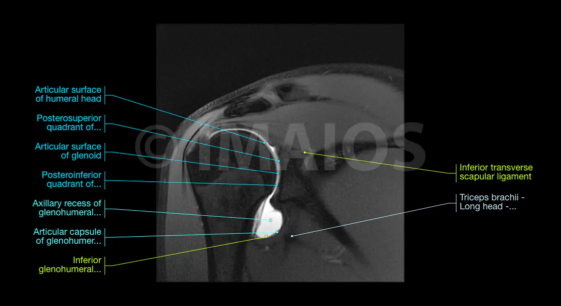
- Oblique coronal PD-weighted fat saturation
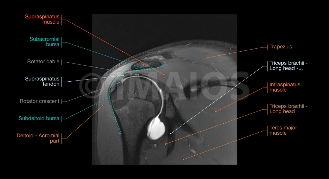
- Oblique sagittal T1-weighted FSE
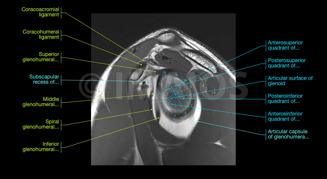
- T1-weighted fat saturation in abduction and external rotation (ABER)
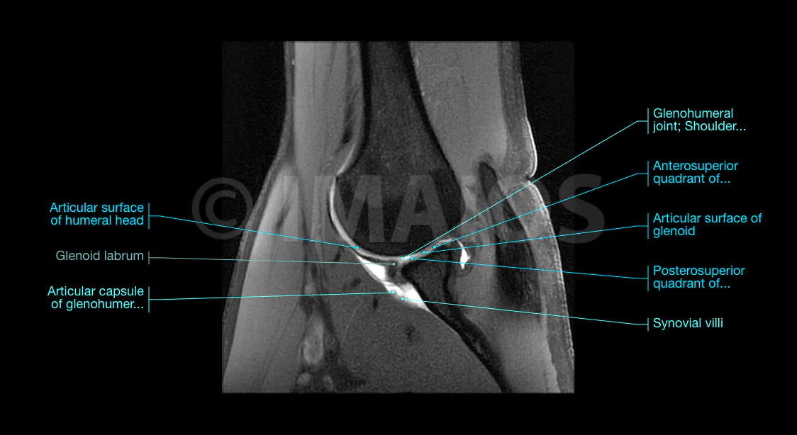
Dr.Antoine Micheau和Dr.Denis Hoa参考解剖学术语(Terminologia Anatomica)2020年最新版,在磁共振图片上标记了解剖结构,并加入常用肩部解剖术语。


- Normal Shoulder MRI and MR Arthrography: Anatomy and Technique. Seminars in musculoskeletal radiology.Llopis, Eva & Montesinos, Pau & Guedez, M & Aguilella, Luis & Cerezal, Luis. (2015). 19. 212-30. 10.1055/s-0035-1549316
- The anterior glenohumeral joint capsule: macroscopic and MRI anatomy of the fasciculus obliquus or so-called ligamentum glenohumerale spirale - Griffin N, Charles-Edwards G, Grant LA. Magnetic resonance cholangiopancreatography: the ABC of MRCP. Insights Imaging. 2012;3(1):11–21. doi:10.1007/s13244-011-0129-9
- Transverse Thickening Along the Articular Surface of the Rotator Cuff Consistent With the Rotator Cable: Identification With MR Arthrography and Relevance in Rotator Cuff Evaluation. Kenneth Sheah, Miriam A. Bredella, Jon J. P. Warner, Elkan F. Halpern, and William E. Palmer American Journal of Roentgenology 2009 193:3, 679-686
- Terminologia anatomica: international anatomical terminology By the Federative Committee on Anatomical Terminology (FCAT). Stuttgart: Georg Thieme Verlag. ISBN-10: 3-13-114361-4. ISBN-13: 978-3-13-114361
这里没有内容

