

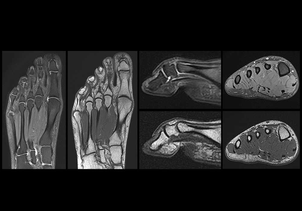
前足正常MRI
前足正常MRI
简介
e-Anatomy这解剖单元专门用于MRI上前足和中足的解剖结构。足分为后足、中足和前足:后足包括距骨和跟骨; 中足由足舟骨、骰骨、内侧、中间和外侧楔骨组成,前足由跖骨和指骨组成。
这解剖单元特别关注前足和脚趾的正常解剖结果,包括骨骼、关节和韧带(跗跖关节、跖趾关节、足底拇趾复合体)、肌腱和肌肉、滑膜腱鞘、足部滑膜囊、前足动脉和静脉,神经供应的详细解剖(足底和背侧神经,足底内侧和外侧神经)。
这足部解剖图谱用于帮助肌肉骨骼放射科医师、风湿病学家和整形外科医生进行日常实践,尤其是用于足底板破裂(草皮脚趾)、籽骨炎、应力性骨折、滑囊炎和其他前足疾病的MR成像。
材料与方法
德国雷根斯堡放射学家Gerald Retzl MD使用Vantage Galan 3T MRI扫描仪对一名健康男性患者进行了前足MR。
使用专用足部线圈在三个解剖平面(轴向平面、矢状平面和冠状平面)上获得了前足的高分辨率MR图像。请注意,足部平面的术语可能会导致识别这些成像平面的混乱,因为定位(俯卧与仰卧)会影响针对身体长轴的相对平面。这是为什么部分作者更喜欢分别使用短轴和长轴术语来表示轴向和冠状面。
虽然我们选择了一名健康的志愿者进行这次MR检查,但我们在标记过程中发现了一些解剖学变化,例如第五脚趾的中节和远端指骨之间的融合,以及拇长屈肌和趾长屈肌腱之间的腱间连接。
选择MRI序列来改善解剖结构(T1加权)和流体(质子密度加权)的呈现。
下面提供了用于该检查的足部成像的MRI参数:
| MRI sequence | Number of images | Slice thickness (mm) | TR (ms) | TE (ms) | FOV (mm) | Matrix | Resolution |
| Axial PD FS | 34 | 2 | 9144 | 40 | 80 | 256x256 | 0.2x0.2x3 |
| Axial T1 | 34 | 2 | 675 | 12 | 80 | 320x256 | 0.2x0.2x3 |
| Sagittal PD FS | 45 | 2 | 9144 | 40 | 80 | 352x256 | 0.2x0.2x3 |
| Sagittal T1 | 45 | 2 | 675 | 12 | 80 | 320x256 | 0.2x0.2x3 |
| Coronal PD FS | 86 | 2 | 9144 | 40 | 80 | 320x256 | 0.2x0.2x3 |
| Coronal T1 | 86 | 2 | 675 | 12 | 80 | 352x256 | 0.2x0.2x3 |
解剖标记是由法国蒙彼利埃放射科医师Antoine Micheau M.D.使用 Terminologia Anatomica 2写上的。
这里没有内容
- High-Resolution MRI of the First Metatarsophalangeal Joint: Gross Anatomy and Injury Characterization - James T. P. D. Hallinan, Sheronda M. Statum, Brady K. Huang, Higor Grando Bezerra, Diego A.L. Garcia, Graeme M. Bydder, and Christine B. Chung RadioGraphics 2020 40:4, 1107-1124
- Edama, M., Takabayashi, T., Yokota, H. et al. Morphological characteristics of the plantar calcaneocuboid ligaments. J Foot Ankle Res 14, 3 (2021). https://doi.org/10.1186/s13047-020-00443-7 -
- Castro M, Melão L, Canella C, Weber M, Negrão P, Trudell D, Resnick D. Lisfranc joint ligamentous complex: MRI with anatomic correlation in cadavers. AJR Am J Roentgenol. 2010 Dec;195(6):W447-55. doi: 10.2214/AJR.10.4674. PMID: 21098178
- Unraveling the plantar plate in MRI: normal and pathologic findings - Alípio Gomes Ormond Filho, Daniel Pastore, Erica Narahashi, Marcelo Astolfi Caetano Nico - Rev ABTPé. 2016;10(2):39-44.
- How to assess plantar plate anatomy and tears on MRI - Atgen C - https://www.youtube.com/watch?v=c5UtS1DRl88
- Great toe MRI Anatomy - First metatarsophalangeal joint- Atgen C - https://www.youtube.com/watch?v=_SuWsLV0Jm4
- https://radiologykey.com/imaging-of-the-forefoot-and-midfoot/
- SIMS - Le pied - http://www.sims-asso.org/uploads/pdfs/monographies/3/1.pdf
- Terminologia anatomica: international anatomical terminology By the Federative Committee on Anatomical Terminology (FCAT). Stuttgart: Georg Thieme Verlag. ISBN-10: 3-13-114361-4. ISBN-13: 978-3-13-114361-7
- Pocket Atlas of Human Anatomy: 5th edition - W. Dauber, Founded by Heinz Feneis
