


Anatomy atlas of the upper limb
Anatomy atlas of the upper limb

This section of the illustrated atlas of human anatomy details the upper limb. 92 anatomical illustrations were created, all in vector format suitable for the web.

This module was designed for medicine students, particularly those preparing for the first year of medical studies (PCEM1), and for health professionals (nursing students, physiotherapy, osteopathy, radiology technicians, physicians, surgeons, radiologists) in orthopedics, rheumatology, functional rehabilitation, radiology...

Illustrations of the anatomy of the upper limb
These images were created using data obtained from medical imaging (CT, MRI, X-rays). Some are 3D views of the bones, vectorised with Adobe Illustrator, while others are diagrams and some are digital paintings produced with Adobe Photoshop.
All of these anatomy illustrations are original and created by Antoine Micheau - MD.

Anatomical structures of the upper limb
630 anatomical structures of the upper limb (pectoral girdle, shoulder, arm, elbow, forearm, wrist, hand and fingers) were labeled.


- The first image shows the different regions of the upper extremity: deltoid region, cubital fossa, thenar eminence and hypothenar...
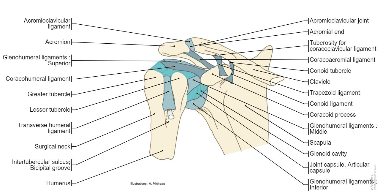
- The next chapter is about the osteology of the upper limb, with all its anatomical structures, muscle insertions and ligaments of the bones: scapula, clavicle, humerus, ulna, radius, carpal bones, metacarpals and phalanges of the fingers.
- The arthrology section includes the study of joints and ligaments forming the upper limb including arcromial-clavicular, sterno-clavicular, radio-ulnar proximal and distal joints, the gleno-humeral, humero-ulnar, carpo-metacarpal, metacarpo-phalangeal and interphalangeal articulations of the hand.
- Multiple illustrations on the myology of the upper limb, with various muscular compartments (fascial compartments), fascia and intermuscular septum, and the muscles and tendons.
- The chapter on the upper limb arteries begins with an overview of the arteries (subclavian, axillary, brachial, radial and ulnar arteries and deep and superficial palmar arches), and then different anatomical images show the arteries of the deltoid region of elbow, hand (palmar and dorsal) and the finger.
- Only the superficial veins of the hand, forearm and arm were represented, the anatomy of the deep veins being similar to the anatomy of the arteries.
- An anatomical diagram illustrates the lymph nodes of the upper limb, with the pectoral, axillary (lymphatic ganglia), paramammary, parasternal, humeral and ulnar nodes.
- The study of the innervation of the upper limb begins with a diagram of the brachial plexus, then the paths of each nerve in the arm and the forearm are detailed (musculocutaneous nerve, median nerve, ulnar nerve, axillary nerve and radial nerve). Finally, the different dermatomes of the nerves of the upper limb are detailed through dorsal and anterior views of the upper limb.
- The final chapter presents anatomical charts of anatomical sections of the upper limb: the axilla and the deltoid region in axial and coronal and axial sections of the arm, the elbow, forearm, wrist, carpal and metacarpal regions.
![Bones of hand - Anatomy : Radius, Ulna, Scaphoid, Lunate, Triquetrum, Pisiform, Trapezium, Trapezoid, Capitate, Hamate, Metacarpals [I -V], Digital bones; Phalanges](https://cdn.imaios.cn/i/images/1/0/4/7/487401-1-eng-GB/upper-limb-illustrations-bones-hand.jpg?auto=format,compress&caption=1&q=75&w=1280&s=c42a9f8979c1f0831eaabc08904fafaf)
Terminology for the study of the anatomy of the upper limb
The anatomical structures were labeled by Antoine Micheau M.D. (Radiologist, Montpellier – France), using the Terminologia Anatomica.

- Terminologia Anatomica: International Anatomical Terminology - FCAT Federative Committee On Anatomical Terminology, Federative Committee on Anatomical Terminology - Thieme, 1998 - ISBN 3131152516, 9783131152510
- Lexique illustré d'anatomie Feneis – Feneis - Editeur : Flammarion Médecine-Sciences (23 août 2007) - Collection : ATLAS DE POCHE - ISBN-10: 225712250X - ISBN-13: 978-2257122506
- Atlas d'anatomie humaine - 4e édition - Frank-H Netter - Pierre Kamina (Traducteur) - Paru le : 25/07/2007 - Editeur : Masson - ISBN : 978-2-294-08042-5 - EAN : 9782294080425 (lien : http://www.netterimages.com/)
- Anatomie topographique, descriptive et fonctionnelle – Alain Bouchet, Jacques Cuilleret – Editions Masson (1991) - Langue : Français - ISBN-10: 2225824673 - ISBN-13: 978-2225824678
- Anatomie, tome 1 (Tronc),2 (Appareil locomoteur), 3 (ORL), 4 (Neuro-anatomie) – Dirigé par Jean-Marc Chevalier - Editeur : Flammarion Médecine (1 novembre 1998) - Collection : Monographies - Langue : Français - ISBN-10: 2257101286 - ISBN-13: 978-2257101280
