

PET-CT atlas of the whole body
PET-CT atlas of the whole body
PET-CT images provided by G. Chuto - MD
This module is about the anatomy of the human body as studied when performing a PET scan with FDG injection.
It contains 280 images in axial section, ranging from cranial base to the root of the thighs, with over 250 anatomical structures labeled, with 4 selectable image types (PET, scanner, or PET-CT)
It is particularly useful for doctors and nuclear medical technicians, radiologists and oncologists, including the disease spread assessment of cancers.
PET-scanner images with FDG
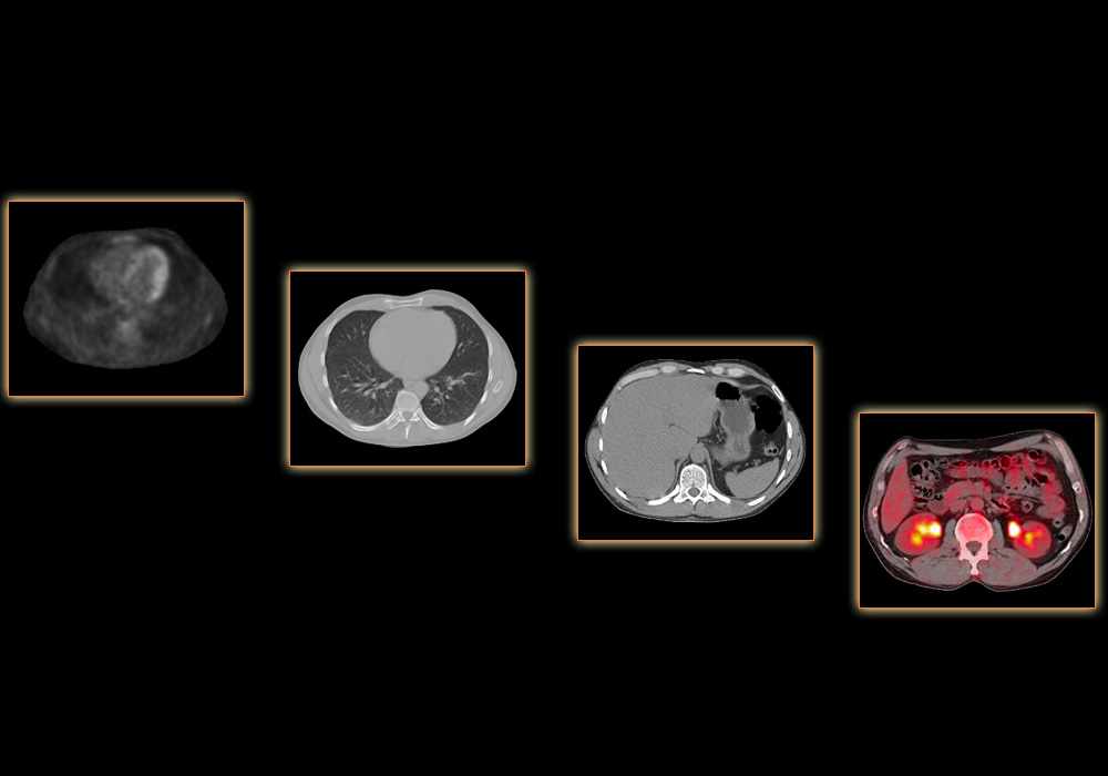
The PET-CT images of a healthy male subject, were provided by G. Chuto - MD. There are four types of images, typically composed of a positron emission tomography combined with a scanner:
- PET-CT fusion: combining image scanner data and FDG tracer scintigraphy. These images are often very educational but are not used on a daily basis.
- CT (abdomen-mediastinum): whole body scanner images without injection with window function that allows the study of the soft tissues, including lymph node structures, mediastinum and abdomen.
- CT (lung-bone): stronger window function, allowing a better view of the pulmonary parenchyma and the lungs.
- PET: tomographic images with attenuation factor correction, allowing to view the areas fixing the tracer (e.g. heart, brain, urinary tract).
Anatomical structures labeled on a PET-CT
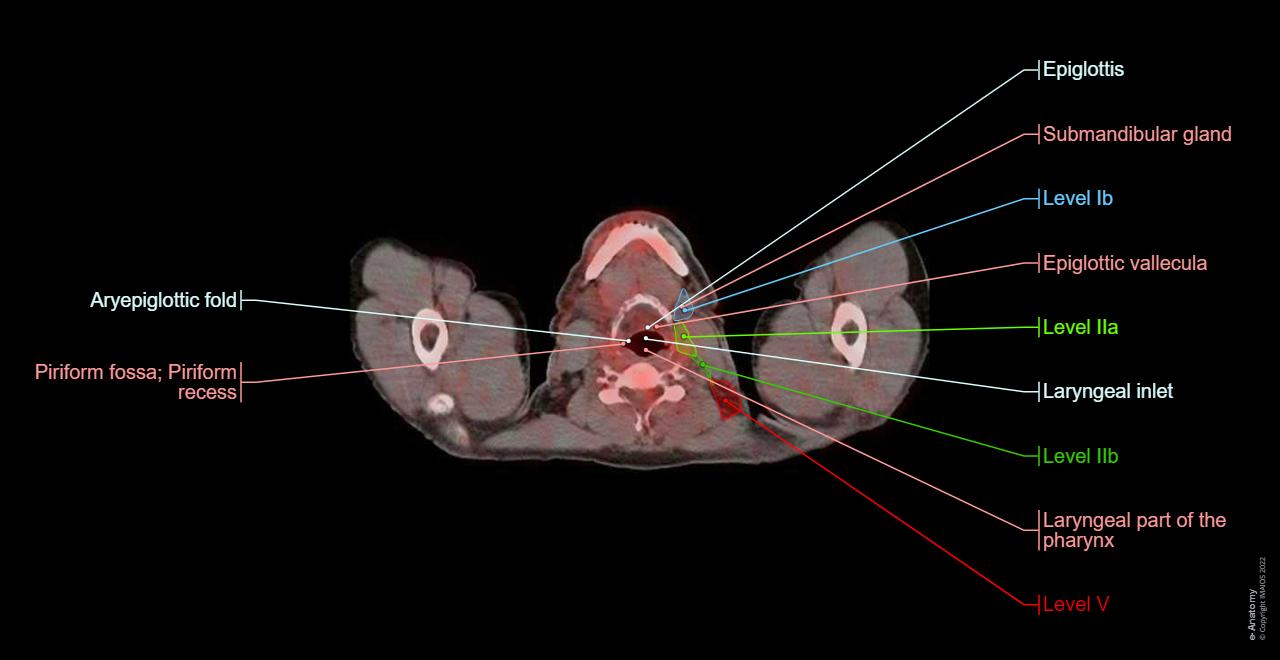
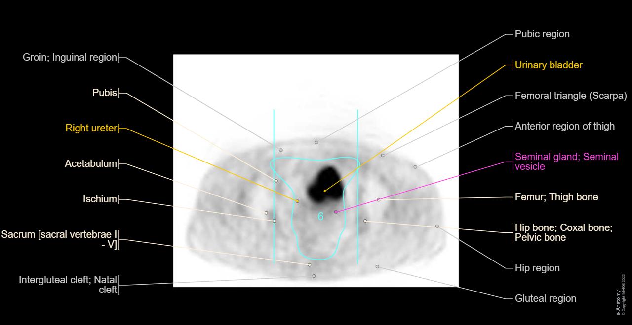
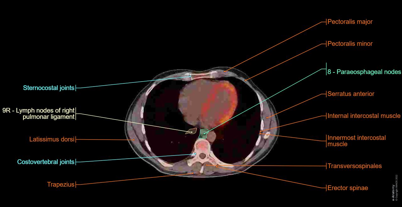
250 anatomical structures of the neck and trunk were labeled using only the visible structures studied through praxis on a PET scanner:
- The general anatomy presents the main regions of human body, useful in locating a tumoral process: axilla, quadrants of the abdomen and etc.
- The bone (skeletal system) roughly presents the different bones of the neck, trunk and root of the lower limbs in order to avoid overloading the module.
- The articular system presents the large joints of the human body, with particular focus on the spine.
- The muscles (muscular system) were exhaustively labeled, apart from the erector spinae muscles and certain muscles of the pelvic floor that are not identifiable on this scanner.
- The respiratory system includes the main structures from the nasal cavity to the bronchi.
- The bronchopulmonary segments were identified by labeled areas with the help of the classification of the Terminologia Anatomica (numbers vary depending on the source areas, e.g. Boyden).
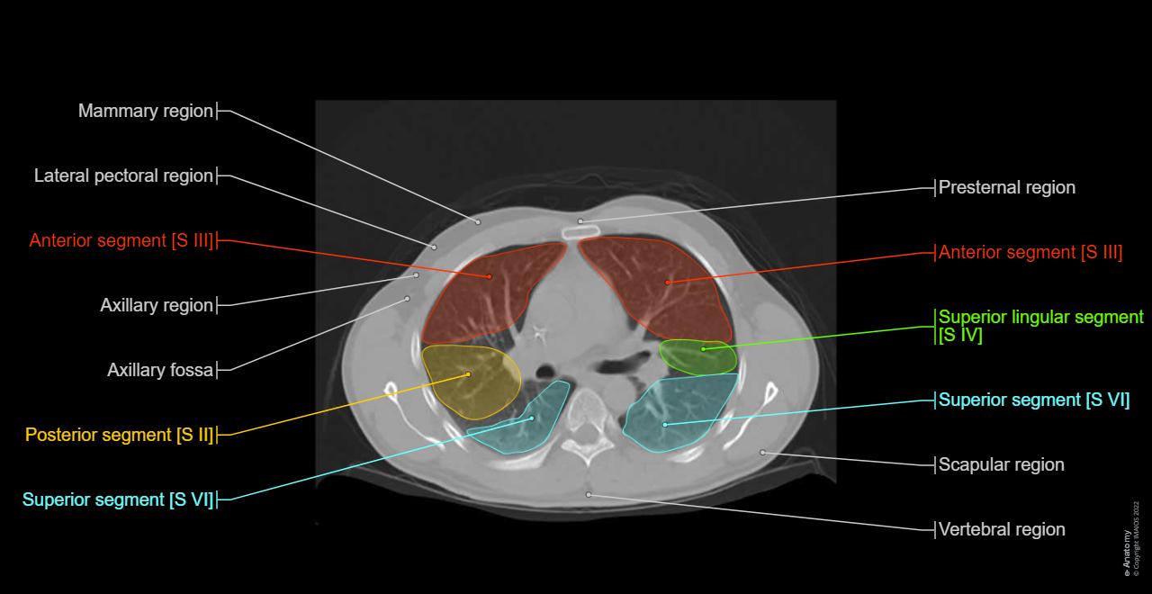
- The thoracic cavity contains the large mediastinal regions and the cardiac structures.
- The digestive system includes the oral cavity, pharynx, oesophagus, stomach, large and small intestines, liver and pancreas. We have included here the spleen, which, however, is a lymphoid and not digestive organ.
- The hepatic segmentation represents the 7 segments of the liver by means of zones and labels.
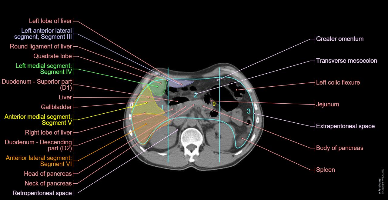
- The abdominal cavity includes the different spaces (peritoneal, retroperitoneal, retropubic) and mesenteries (mesentery, mesocolon).
- The Peritoneal Cancer Index is presented in the form of zones numbered 1 through 12 of the different possible locations of peritoneal carcinomatosis. A diagram of this index is available in the classifications tab. This index is still not officially recognized but widely used in clinical practice.
- The urinary system is represented mainly by the kidneys and bladder.
- The male reproductive system was labeled in a simple way to quickly locate structures such as the seminal vesicles or the prostate.
- The endocrine glands include the thyroid, the thymus (illustrated despite its atrophying into adulthood) and the adrenal glands.
- The arteries were superficially labeled, primarily as they form basic anatomical landmarks.
- The veins include the upper and lower vena cava system as well as the portal system.
- The lymph nodes tab describes the lymph nodes as described in the Terminologia Anatomica – names often not used on a daily practice.
- The ganglionic areas pick up the classifications used in oncology and surgery (cervical and thoracic lymph nodes (IASLC)) in the form of differently colored regions.
- The nervous system is not easy to study on a CT scan without injection.
Anatomical terminology
The anatomical structures were labeled by Antoine Micheau M.D. (Radiologist, Montpellier – France), using the Terminologia Anatomica.
No content available
