Measurements of axes and angles in canine orthopedics
Measurements of axes and angles in canine orthopedics
Introduction
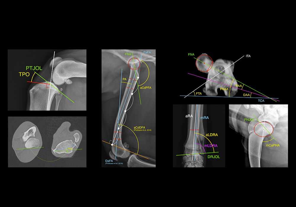
The graphic representation of bone axes and joint angles provides an in-depth understanding of the biomechanics and pathological processes that can affect the thoracic and pelvic limbs in dogs. The use of precise, reproducible measurements guarantees reliable, comparable values within an animal and from one study to the other.
These measurements have been particularly refined for understanding angular deformities of the long bones, coxofemoral and elbow dysplasia, congenital luxation of the patella and rupture of the cranial cruciate ligament. They are also an essential step in planning corrective (surgical) procedures and evaluating their results.
In this module, we have grouped together the main radiology measurements described in canine orthopedics, including pelvimetry. The anatomical landmarks required for these measurements have been highlighted.

Material and methods
Radiographic exams and CT scans of the thoracic and pelvic limbs of dogs were provided by Dr Susanne Boroffka, DVM, dipl. ECVDI, PhD (Utrecht, Netherlands) and Thierry Boulet, DVM, Certificate of Advanced Education in Animal Osteoarticular Traumatology and Orthopedics (St Jean de Védas, France).
Measures were drawn on the images by Stephan Mahler DVM, MA, MSc, PhD (Veterinary Anatomist - IMAIOS). They have been grouped into different themes:
- Bones
- Bony landmarks
- Bone axes
- Joint orientation lines
- Joint orientation angles
- Bone measurements
- Bone areas
- Percentages and indexes
Definitions of the measures were systematically proposed. Normal values of the anatomical measurements found in the literature were supplemented.
List of abbreviations:
a-mAA: Anatomical-mechanical axis angle
AAA: Acetabular anteversion angle
aCdDFA: Anatomic caudal distal femoral angle
aCdDRA: Anatomic caudal distal radial angle
aCdPFA: Anatomic caudal proximal femoral angle
aCdPRA: Anatomic caudal proximal radial angle
aCrPRA: Anatomic cranial proximal radial angle
AD: Acetabular depth
aFA: Anatomic femoral axis
aHA: Anatomic humeral axis
AI: Acetabular index
AIA: Acetabular index angle
aLDFA: Anatomic lateral distal femoral angle
aLDHA: Anatomic lateral distal humeral angle
aLDRA: Anatomic lateral distal radial angle
aLPFA: Anatomic lateral proximal femoral angle
aMPHA: Anatomic medial proximal humeral angle
aMPRA: Anatomic medial proximal radial angle
aRA: Anatomic radial axis
aTA: Anatomical tibial axis
AW: Acetabular width
BAD: Bi-acetabular distance
BID: Bi-iliac distance
CD: Conjugata diagonalis
CdCTA: Caudal condylar tibial axis
CEA: Center-edge angle
DAA: Distal angle of anteversion
DaFA: Distal anatomic femoral axis
DASA: Dorsal acetabular sector angle
DCrTA: Distal cranial tibial axis
DFHCAI: Dorsal femoral head coverage area index
DFHCWI: Dorsal femoral head coverage width index
DFJOL: Distal femoral joint orientation line
DHJOL: Distal humeral joint orientation line
DPTA: Diaphyseal proximal tibial angle
DRJOL: Distal radial joint orientation line
DTJOL: Distal tibial joint orientation line
ECA: Elbow compression angle
ERP: Elbow rotational position
FHCP: Femoral head coverage percentage
FIA: Femoral inclination angle
FNA: Femoral neck axis
FNAA: Femoral neck anteversion angle
FPA: Femoral procurvation angle
FTA: Femoral trochanteric angle
FVA: Femoral varus angle
GTI: Greater tubercle inclination
GTO: Greater tubercle offset
HASA: Horizontal acetabular sector angle
HTA: Humeral torsion angle
HTEA: Horizontal toit externe angle
HTED: Humeral transepicondylar distance
IA: Iliac axis
ID: Distraction index
LBID: Lateral bi-ischiatic distance
MBID: Medial bi-ischiatic distance
mCaPHA: Mechanical caudal proximal humeral angle
mCdDFA: Mechanical caudal distal femoral angle
mCdDTA: Mechanical caudal distal tibial angle
mCdPFA: Mechanical caudal proximal femoral angle
mCdPTA: Mechanical caudal proximal tibial angle
mCMA: Mechanical carpus/metacarpus axis
mCrDHA: Mechanical cranial distal humeral angle
mCrDTA: Mechanical cranial distal tibial angle
mCrPTA: Mechanical cranial proximal tibial angle
mFA: Mechanical femoral axis
mHA: Mechanical humeral axis
mLDFA: Mechanical lateral distal femoral angle
mLDHA: Mechanical lateral distal humeral angle
mLDRA: Mechanical lateral distal radial angle
mLDTA: Mechanical lateral distal tibial angle
mLPCMA: Mechanical lateral proximal carpus/metacarpus angle
mLPFA: Mechanical lateral proximal femoral angle
mLPTA: Mechanical lateral proximal tibial angle
mMDTA: Mechanical medial distal tibial angle
mMPHA: Mechanical medial proximal humeral angle
mMPRA: Mechanical medial proximal radial angle
mMPTA: Mechanical medial proximal tibial angle
mRA: Mechanical radial axis
mTA: Mechanical tibial axis
NA: Norberg angle
PAA: Proximal angle of anteversion
PaFA: Proximal anatomic femoral axis
PFJOL: Proximal femoral joint orientation line
PHJOL: Proximal humeral joint orientation line
PRJOL: Proximal radial joint orientation line
PTA: Patellar tendon angle
PTA: Proximal tibial axis
PTJOL: Proximal tibial joint orientation line
PTTA: Proximal tibial tuberosity angle
QA: Quadriceps angle
RTA: Radial torsion angle
rTTW: Relative tibial tuberosity width
SD: Diameter sagittalis
TCTA: Transcondylar tibial axis
TPA: Tibial plateau angle
TPO: Tibial plateau orientation
TTA: Tibial torsion angle
TV: Tibial valgus
VASA: Ventral acetabular sector angle
ZA: Z angle
θ: θ angle
Images


- Aertsens A, Rincon Alvarez J, Poncet CM, Beaufrère H, Ragetly GR. Comparison of the tibia plateau angle between small and large dogs with cranial cruciate ligament disease. Vet Comp Orthop Traumatol. 2015;28(6):385-90. doi: 10.3415/VCOT-14-12-0180. Epub 2015 Sep 18.
- Aghapour M, Bockstahler B, Vidoni B. Evaluation of the Femoral and Tibial Alignments in Dogs: A Systematic Review. Animals (Basel). 2021 Jun 17;11(6):1804. doi: 10.3390/ani11061804. PMID: 34204283; PMCID: PMC8234394.
- Al Aiyan A, Richardson K, Manchi G, Ginja M, Brunnberg L. Measurement of the Femoral Anteversion Angle in Medium and Large Dog Breeds Using Computed Tomography. Front Vet Sci. 2021 Mar 5;8:540406. doi: 10.3389/fvets.2021.540406
- Andronescu et al. (2015). Associations between early radiographic and computed tomographic measures and canine hip joint osteoarthritis at maturity. American Journal of Veterinary Research, 76(1), 19–27. doi:10.2460/ajvr.76.1.19
- Aper R, Kowaleski MP, Apelt D, Drost WT, Dyce J. Computed tomographic determination of tibial torsion in the dog. Vet Radiol Ultrasound. 2005 May-Jun;46(3):187-91. doi: 10.1111/j.1740-8261.2005.00048.x.
- Baroni E, Matthias RR, Marcellin-Little DJ, Vezzoni A, Stebbins ME. Comparison of radiographic assessments of the tibial plateau slope in dogs. Am J Vet Res. 2003 May;64(5):586-9. doi: 10.2460/ajvr.2003.64.586. PMID: 12755299
- Belkoff SM, Padgett G, Soutas-Little RW. Development of a device to measure canine coxofemoral joint laxity. Vet Comp Orthop Traumatol 1989, 2, 31-36.
- Comhaire et al. Canine hip dyslasia: the significance of the Norberg angle for healthy breeding. Journal of Small Animal Practice (2011) 52, 536–542.DOI: 10.1111/j.1748-5827.2011.01105.x
- Dismukes DI, Tomlinson JL, Fox DB, Cook JL, Witsberger TH. Radiographic measurement of canine tibial angles in the sagittal plane. Vet Surg. 2008 Apr;37(3):300-5. doi: 10.1111/j.1532-950X.2008.00381.x.
- Dobak, Tetyda P.; Voorhout, George; Vernooij, Johannes C.M.; Boroffka, Susanne A.E.B. (2018). Computed tomographic pelvimetry in English bulldogs. Theriogenology, (), S0093691X18302437–. doi:10.1016/j.theriogenology.2018.05.025
- Dudley RM, Kowaleski MP, Drost WT, Dyce J. Radiographic and computed tomographic determination of femoral varus and torsion in the dog. Vet Radiol Ultrasound. 2006 Oct-Nov;47(6):546-52. doi: 10.1111/j.1740-8261.2006.00184.x.
- Duncan et al. (2022). Assessment of normal radial joint orientation angles in nonchondrodystrophic small-breed dogs. J Am Vet Med Assoc. Sep 1;261(1):1-4. doi: 10.2460/javma.22.07.0286.
- Eneroth et al. (1999). Radiographic pelvimetry for assessment of dystocia in bitches: a clinical studv in two terrier breeds. Journal of Small Anmal Practice. J Small Anim Pract;40.257-264. DOI: 10.1111/j.1748-5827.1999.tb03076.x
- Fox J, Tomlinson JL. Principles of Angular Limb Deformity Correction. In: Tobias KM, Johnston SA, editors. Veterinary Surgery Small Animal. St Louis (MI): Elsevier Saunders; 2012. p.657-668.
- Fox et al. (2006). Principles of Uniapical and Biapical Radial Deformity Correction Using Dome Osteotomies and the Center of Rotation of Angulation Methodology in Dogs. Veterinary Surgery;35:67–77. doi:10.1111/j.1532-950X.2005.00114.x
- Garnoeva, R, Roydev, R, Paskalev, M et al. Radiographic measures of pelvic limb malalignment in small breed dogs with various grades of medial patellar luxation. Comp Clin Pathol 2018;27, 1551–1555. doi.org/10.1007/s00580-018-2772-8.
- Ginja et al. (2007). Measurement of the femoral neck anteversion angle in the dog using computed tomography.174(2), 378–383. doi:10.1016/j.tvjl.2006.08.002
- Goodrich et al. Thoracic Limb Alignment in Healthy Labrador Retrievers: Evaluation of Standing Versus Recumbent Frontal Plane Radiography. Veterinary Surgery 43 (2014) 791–803. DOI:10.1111/j.1532-950X.2014.12140.x
- Guénégo L, Payot M, Charru P, Verwaerde P. Comparison of tibial anatomical-mechanical axis angle between predisposed dogs and dogs at low risk for cranial cruciate ligament rupture. Vet J. 2017 Jul;225:35-41. doi: 10.1016/j.tvjl.2017.04.011. Epub 2017 May 3. PMID: 28720297.
- Hauptman J, Prieur WD, Butler HC, Guffy MM. The angles of inclination of the canine femoral head and neck. Vet Surg. 1979;8(3):74-77. doi.org/10.1111/j.1532-950X.1979.tb00612.x
- Horňáková et al. (2023). Radiographic pelvimetry in relation to dystocia in Bulldogs. Folia Veterinaria 67;3:33-38. DOI: 10.2478/fv-2023-0025
- Inauen R, Koch D, Bass M, Haessig M. Tibial tuberosity conformation as a risk factor for cranial cruciate ligament rupture in the dog. Vet Comp Orthop Traumatol. 2009;22(1):16-20. PMID: 19151865.
- Janovec J, Kyllar M, Midgley D, Owen M. Conformation of the proximal tibia and cranial cruciate ligament disease in small breed dogs. Vet Comp Orthop Traumatol. 2017 May 22;30(3):178-183. doi: 10.3415/VCOT-16-07-0115. Epub 2017 Mar 23.
- Jeong J et al. (2020) Measurement of Thoracic Limb Joint Reference Angles in Purebred Shih-Tzu Dogs by Computed Tomography. J Vet Clin 37(4):169 doi.org/10.17555/jvc.2020.08.37.4.169
- Kara ME, Sevil-Kilimci F, Dilek ÖG, Onar V. Proximal and distal alignment of normal canine femurs: A morphometric analysis. Ann Anat. 2018 May;217:125-128. doi: 10.1016/j.aanat.2018.02.006. Epub 2018 Mar 19.
- Kim SE, Lewis DD. Corrective osteotomy for procurvatum deformity caused by distal femoral physeal fracture malunion stabilised with String-of-Pearls locking plates: results in two dogs and a review of the literature. Aust Vet J. 2014 Mar;92(3):75-80. doi: 10.1111/avj.12149.
- Kroner et al. (2017). Assessment of radial torsion using computed tomography in dogs with and without antebrachial limb deformity. Veterinary Surgery, 46(1), 24–31. doi:10.1111/vsu.12589
- Kwan et al (2014). Correction of Biapical Radial Deformities by Use of Bi-Level Hinged Circular External Fixation and Distraction Osteogenesis in 13 Dogs. Veterinary Surgery, 43(3), 316–329. doi:10.1111/j.1532-950X.2014.12114.x
- Kwon et al. (2022). Evaluation of the radial procurvatum using the center of rotation of angulation methodology in chondrodystrophic. Dogs Front Vet Sci; 8:774993. doi: 10.3389/fvets.2021.774993
- Lopez et al. (2008) Relationships among measurements obtained by use of computed tomography and radiography and scores of cartilage microdamage in hip joints with moderate to severe joint laxity of adult dogs. Am J Vet Res. Mar;69(3):362-70. doi: 10.2460/ajvr.69.3.362.
- Lusetti F, Bonardi A, Eid C, Brandstetter de Belesini A, Martini FM. Pelvic limb alignment measured by computed tomography in purebred English Bulldogs with medial patellar luxation. Vet Comp Orthop Traumatol. 2017 May 10;30(3):200-208. doi: 10.3415/VCOT-16-07-0116. Epub 2017 May 5.
- Meola et al. (2008). Validation of a Technique to Assess Radial Torsion in the Presence of Procurvatum and Valgus Deformity Using Computed Tomography: A Cadaveric Study. , 37(6), 525–529. doi:10.1111/j.1532-950x.2008.00399.x
- Montavon PM, Hohn RB, Olmstead ML, Rudy RL. Inclination and Anteversion Angles of the Femoral Head and Neck in the Dog Evaluation of a Standard Method of Measurement. Vet Surg. 1985 14(4), 277–82. doi:10.1111/j.1532-950x.1985.tb00883.x
- Mostafa AA, Cunningham DP, Boudrieau RJ, Kowaleski MP, Griffon DJ. Influence of radiographic techniques on the measurement of femoral anteversion angles and a conformation score of pelvic limbs in Labrador retrievers. Vet Surg. 2018 Apr;47(3):421-430. doi: 10.1111/vsu.12782. Epub 2018 Mar 12.
- Mostafa AA, Griffon DJ, Thomas MW, Constable PD. Morphometric characteristics of the pelvic limbs of Labrador Retrievers with and without cranial cruciate ligament deficiency. Am J Vet Res. 2009 Apr;70(4):498-507. doi: 10
- Mostafa et al. (2022) Quantitative assessment of hip morphology to enhance the identification of hip dysplasia in German Shepherd Dogs. Am J Vet Res. 2023 Feb 7;84(3):ajvr.22.09.0165. doi: 10.2460/ajvr.22.09.0165
- Mostafa et al (2022) Modified FCI (Fédération Cynologique Internationale) Scoring of the Coxofemoral Joint in Labrador Retrievers Without and With Hip Dysplasia. Front. Vet. Sci. 9:800237. doi: 10.3389/fvets.2022.800237
- Newman M, Voss K. Computed tomographic evaluation of femoral and tibial conformation in English Staffordshire Bull Terriers with and without congenital medial patellar luxation. Vet Comp Orthop Traumatol. 2017 May 22;30(3):191-199. doi: 10.3415/VCOT-16-12-0162. Epub 2017 Mar 23. Erratum in: Vet Comp Orthop Traumatol. 2017 Jul 10;30(4)
- Olimpo M, Piras LA, Peirone B. Pelvic limb alignment in small breed dogs: a comparison between affected and free subjects from medial patellar luxation. Vet Ital. 2016 Jan-Mar;52(1):45-50. doi: 10.12834/VetIt.71.206.3.
- Osmond CS, Marcellin-Little DJ, Harrysson OL, Kidd LB. Morphometric assessment of the proximal portion of the tibia in dogs with and without cranial cruciate ligament rupture. Vet Radiol Ultrasound. 2006 Mar-Apr;47(2):136-41. doi: 10.1111/j.1740-8261.2006.00119.x. PMID: 16553144.
- Peterson JL, Torres BT, Hutcheson KD, Fox DB. Radiographic determination of normal canine femoral alignment in the sagittal plane: A cadaveric pilot study. Vet Surg. 2020 Aug;49(6):1230-1238. doi: 10.1111/vsu.13465. Epub 2020 Jun 2.
- Phetkaew T, Kalpravidh M, Penchome R, Wangdee C. A Comparison of Angular Values of the Pelvic Limb with Normal and Medial Patellar Luxation Stifles in Chihuahua Dogs Using Radiography and Computed Tomography. Vet Comp Orthop Traumatol. 2018 Feb;31(2):114-123. doi: 10.3415/VCOT-17-05-0067. Epub 2018 Mar 13. Erratum in: Vet Comp Orthop Traumatol. 2018 Feb;31(2):a1.
- Pinna S, Pizzuti E, Carli F. Effects of intertrochanteric varus osteotomy on Norberg angle and percent coverage of the femoral head in displastic dogs. J Vet Sci. 2013;14(2):185-91. doi: 10.4142/jvs.2013.14.2.185.
- Powers MY, Karbe GT, Gregor TP, McKelvie P, Culp WT, Fordyce HH, Smith GK. Evaluation of the relationship between Orthopedic Foundation for Animals' hip joint scores and PennHIP distraction index values in dogs. J Am Vet Med Assoc. 2010 Sep 1;237(5):532-41. doi: 10.2460/javma.237.5.532. PMID: 20807130.
- Puerto DA, Smith GK, Gregor TP, LaFond E, Conzemius MG, Cabell LW, McKelvie PJ. Relationships between results of the Ortolani method of hip joint palpation and distraction index, Norberg angle, and hip score in dogs. J Am Vet Med Assoc. 1999 Feb 15;214(4):497-501.
- Ragetly CA, Evans R, Mostafa AA, Griffon DJ. Multivariate analysis of morphometric characteristics to evaluate risk factors for cranial cruciate ligament deficiency in Labrador retrievers. Vet Surg. 2011 Apr;40(3):327-33. doi: 10.1111/j.1532-950X.2010.00787.x. Epub 2011 Feb 11.
- Rumph PF, Hathcock JT. A symmetric axis-based method for measuring the projected femoral angle of inclination in dogs. Vet Surg. 1990 Sep-Oct;19(5):328-33. doi: 10.1111/j.1532-950x.1990.tb01200.x.
- Sarierler, M. Comparison of femoral inclination angle measurements in dysplastic and nondysplastic dogs of different breeds. Acta Vet. 2004, 52, 245–252.doi: https://doi.org/10.1556/avet.52.2004.2.13
- Schachner and Lopez (2015) Diagnosis, prevention, and management of canine hip dysplasia: a review. Vet Med (Auckl). May 19;6:181-192. doi: 10.2147/VMRR.S53266.
- Schwandt CS, Bohorquez-Vanelli A, Tepic S, Hassig M, Dennler R, Vezzoni A, Montavon PM. Angle between the patellar ligament and tibial plateau in dogs with partial rupture of the cranial cruciate ligament. Am J Vet Res. 2006 Nov;67(11):1855-60. doi: 10.2460/ajvr.67.11.1855. PMID: 17078746.
- Smith et al. Three-dimensional assessment of curvature, torsion, and canal flare index of the humerus of skeletally mature nonchondrodystrophic dogs. Am J Vet Res 2017;78:1140–1149 https://doi.org/10.2460/ajvr.78.10.1140
- Slocum B, Slocum TD. Tibial plateau leveling osteotomy for repair of cranial cruciate ligament rupture in the canine. Vet Clin North Am Small Anim Pract 1993; 23: 777–795.
- Soparat C, Wangdee C, Chuthatep S, Kalpravidh M. Radiographic measurement for femoral varus in Pomeranian dogs with and without medial patellar luxation. Vet Comp Orthop Traumatol. 2012;25(3):197-201. doi: 10.3415/VCOT-11-04-0057. Epub 2012 Jan 27.
- Su L, Townsend KL, Au J, Wittum TE. Comparison of tibial plateau angles in small and large breed dogs. Can Vet J. 2015 Jun;56(6):610-4. PMID: 26028684; PMCID: PMC4431160.
- Tomlinson J, Fox D, Cook JL, Keller GG. Measurement of femoral angles in four dog breeds. Vet Surg. 2007 Aug;36(6):593-8. doi: 10.1111/j.1532-950X.2007.00309.x.
- Vedrine B, Guillemot A, Fontaine D, Ragetly GR, Etchepareborde S. Comparative anatomy of the proximal tibia in healthy Labrador Retrievers and Yorkshire Terriers. Vet Comp Orthop Traumatol. 2013;26(4):266-70. doi: 10.3415/VCOT-12-02-0018. Epub 2013 Apr 24.
- Witte PG. Tibial anatomy in normal small breed dogs including anisometry of various extracapsular stabilizing suture attachment sites. Vet Comp Orthop Traumatol. 2015;28(5):331-8. doi: 10.3415/VCOT-14-12-0186. Epub 2015 Jul 21.
- Wood et al. (2014). Determination of the Mechanical Axis and Joint Orientation Lines in the Canine Humerus: A Radiographic Cadaveric Study. Veterinary Surgery, 43(4), 414–417. doi:10.1111/j.1532-950x.2014.12134.x
- Yasukawa S, Edamura K, Tanegashima K, Seki M, Teshima K, Asano K, Nakayama T, Hayashi K. Evaluation of bone deformities of the femur, tibia, and patella in Toy Poodles with medial patellar luxation using computed tomography. Vet Comp Orthop Traumatol. 2016;29(1):29-38. doi: 10.3415/VCOT-15-05-0089. Epub 2015 Dec 7.
- Žilinčík M, Hluchý M, Takáč L, Ledecký V. Comparison of Radiographic Measurements of the Femur in Yorkshire Terriers with and without Medial Patellar Luxation. Vet Comp Orthop Traumatol. 2018 Jan;31(1):17-22. doi: 10.3415/VCOT-17-01-0018. Epub 2018 Jan 11.