

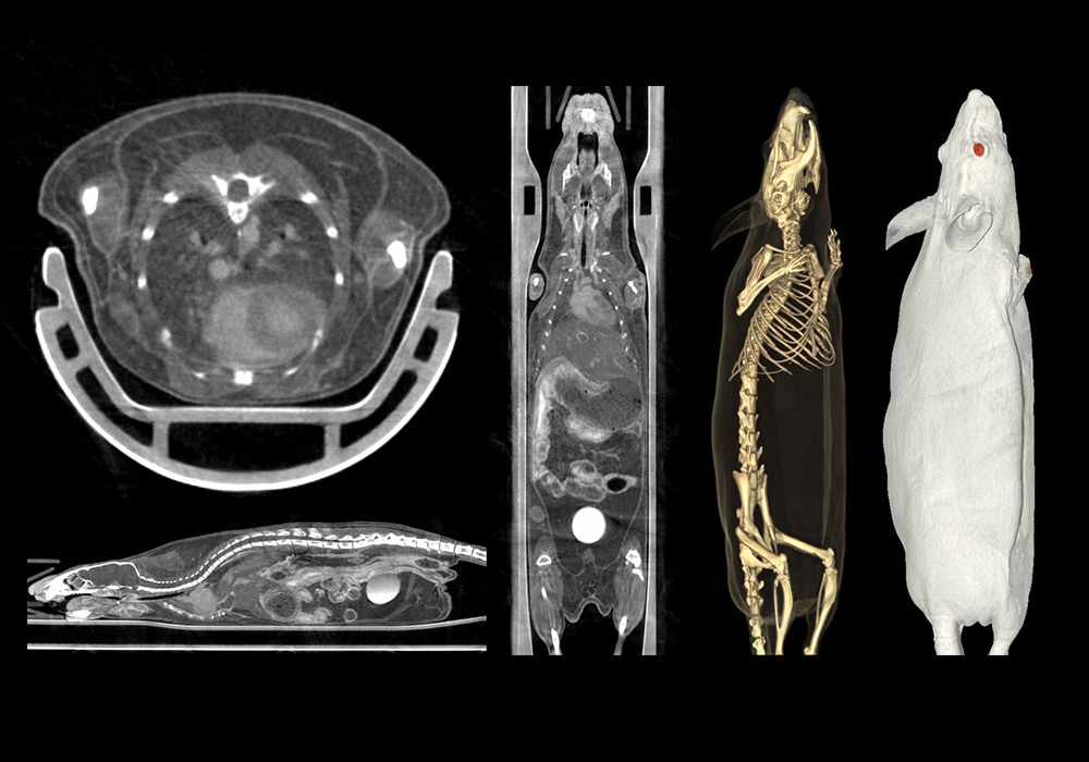
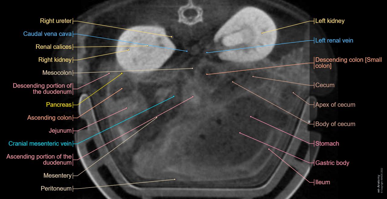
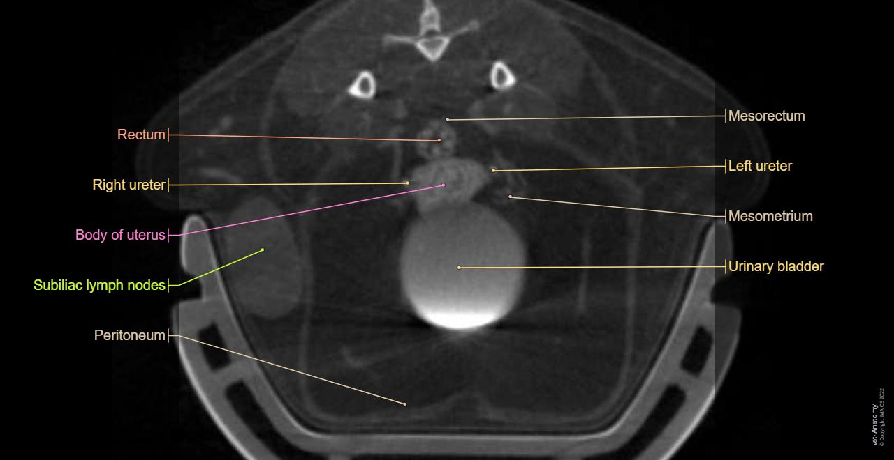
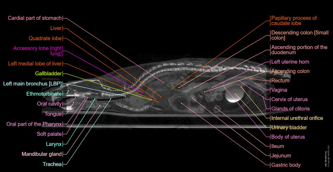
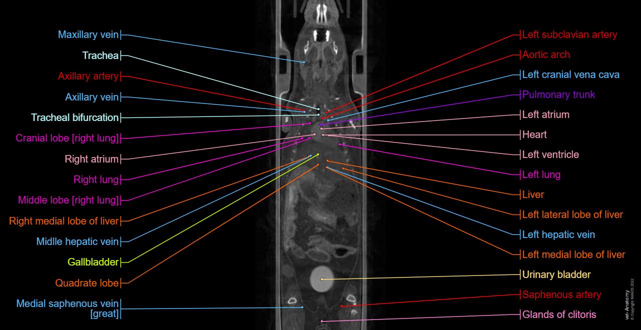
Labeled cross-sectional anatomy of the mouse on micro-CT
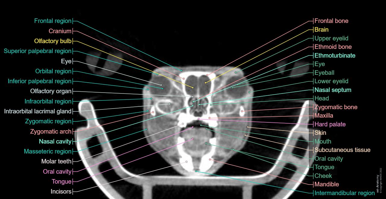
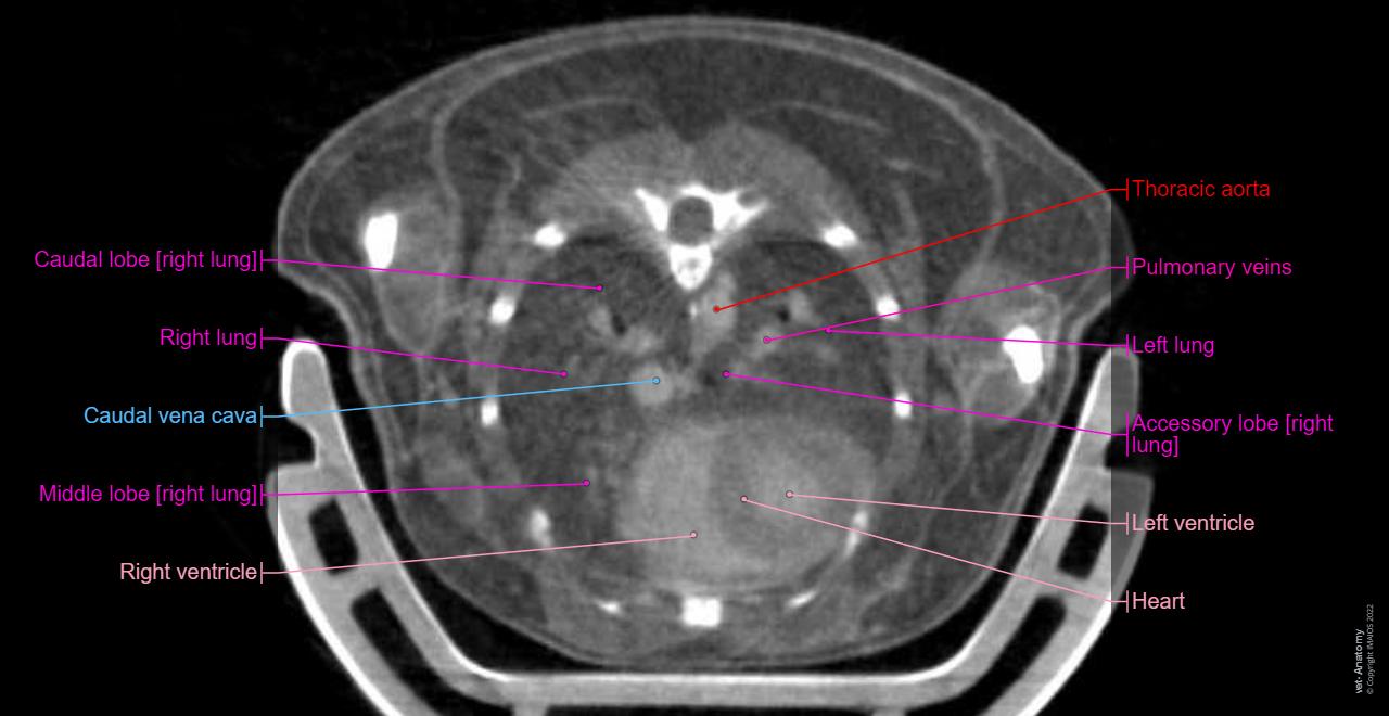
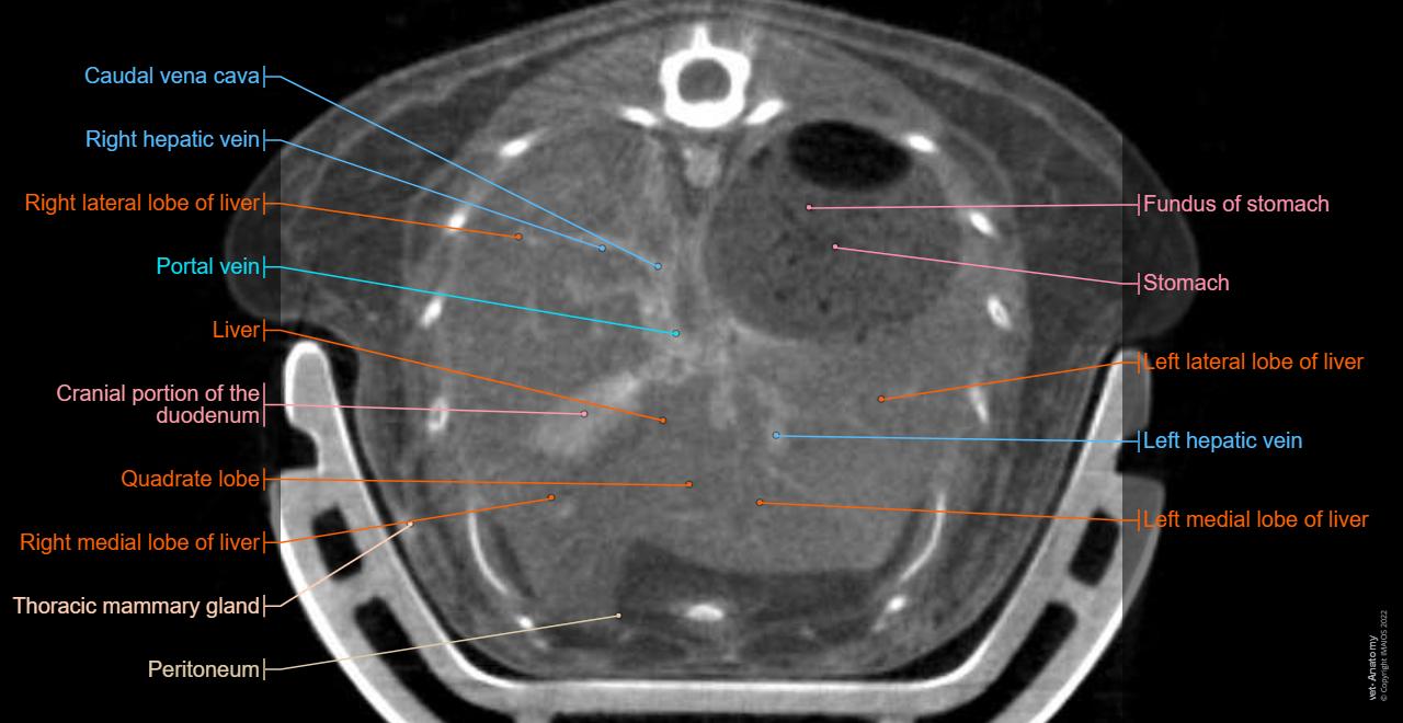
Labeled cross-sectional anatomy of the mouse on micro-CT
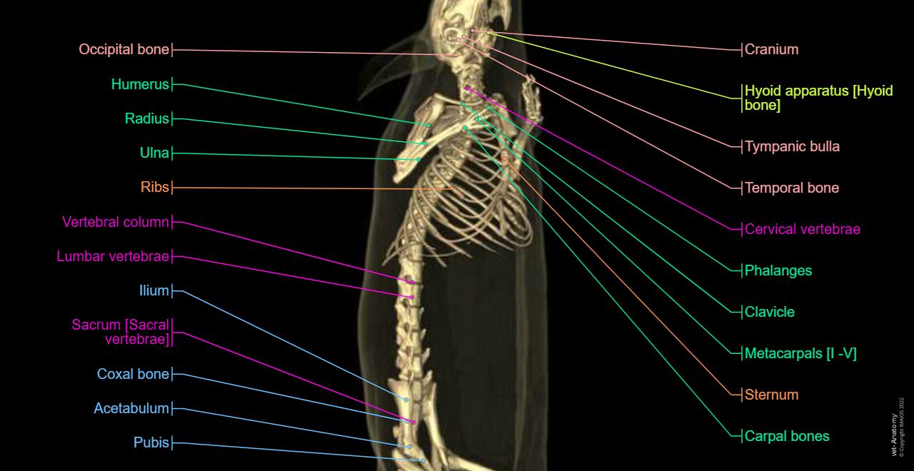
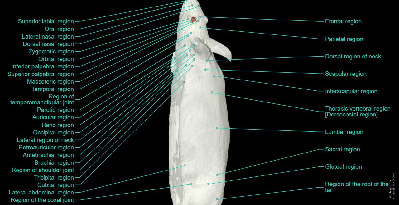
These images of a normal female Swiss mouse have been acquired with a laboratory-based microCT system (nanoScan PET/CT Mediso (Budapest, Hungary) with an operation voltage of 50kVp and a 0,14mm pitch, with an intravenous injection of 2ml of Visipaque (320mg d’I/ml), at CERIMED (Centre Européen de Recherche en Imagerie Médicale (CERIMED), Université d’Aix-Marseille, Marseille, France).
Images were post processed on Osirix Dicom viewer with transverse, sagittal and dorsal reconstructions.
3D rendered images (bones and skin) were created on VGSTUDIOMAX by the Dr. Samuel Mérigeaud (Tridilogy, Montpellier, France).
No content available