

Atlas of feline anatomy on X-ray images
Atlas of feline anatomy on X-ray images
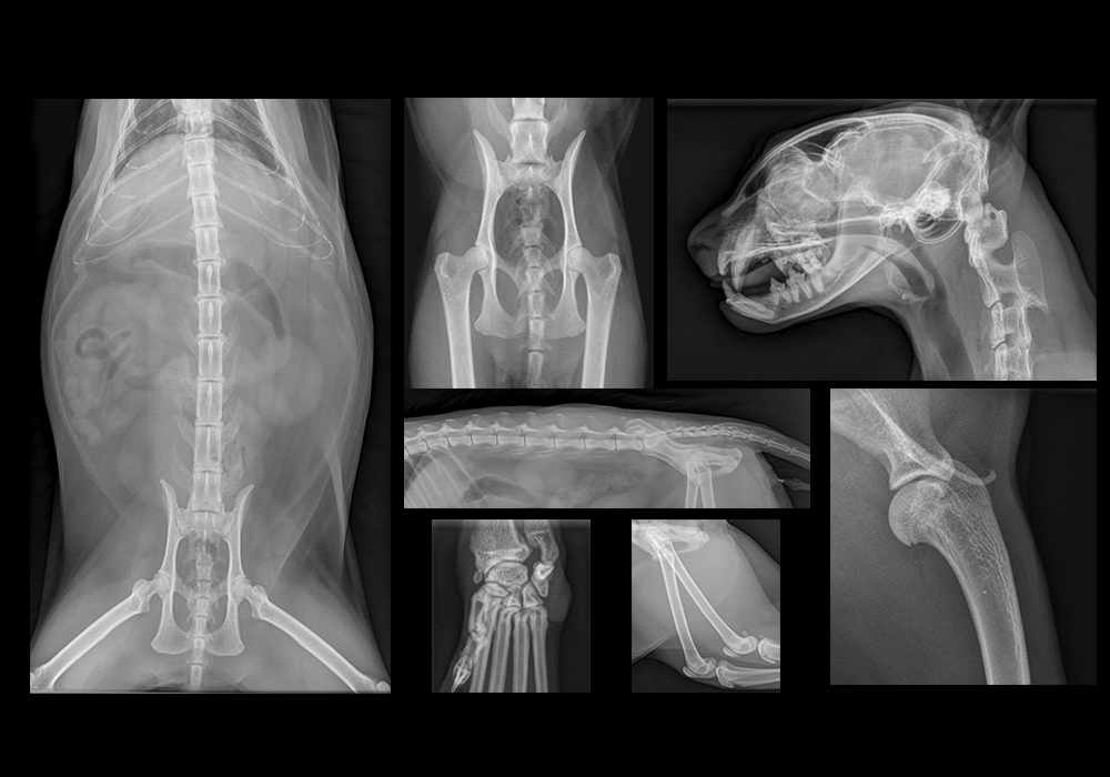
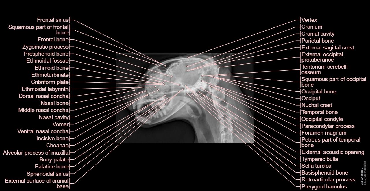
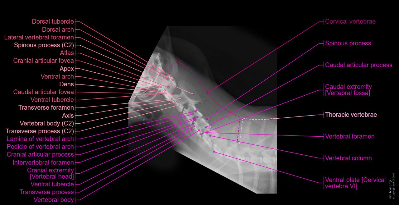
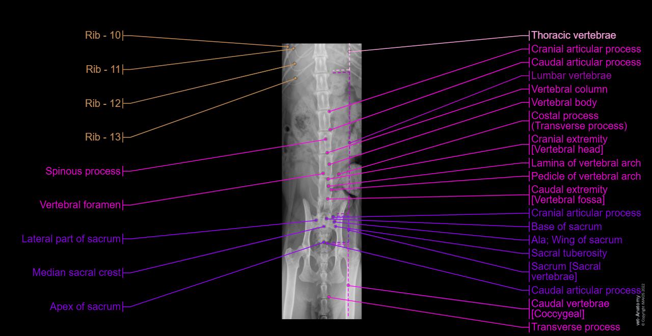
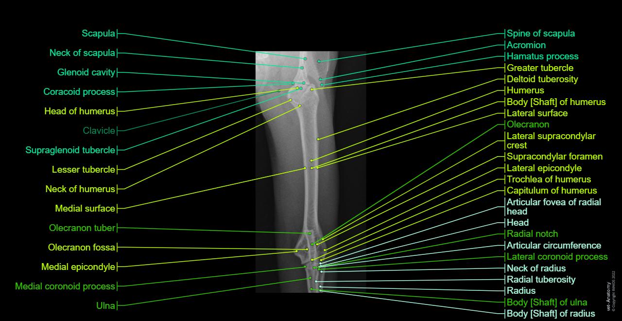
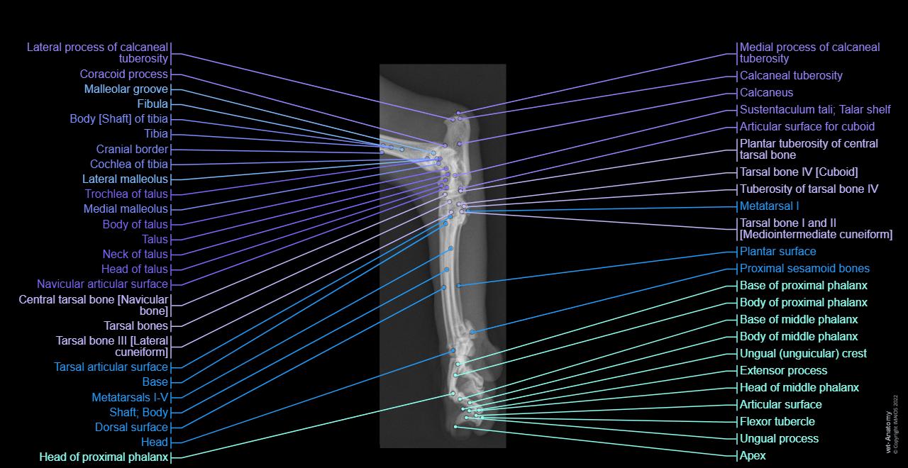
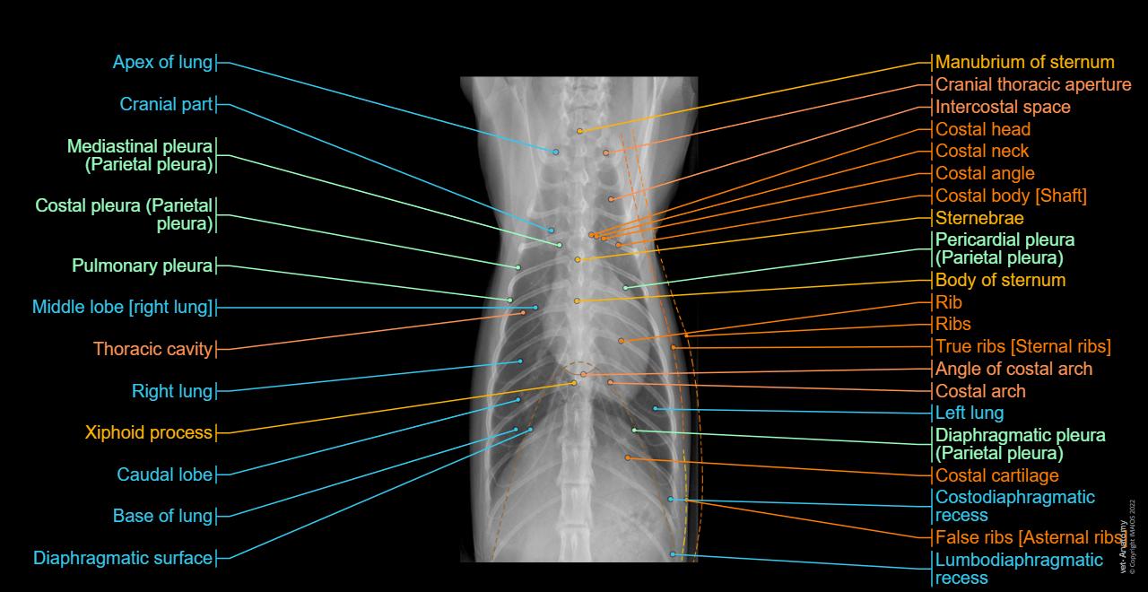
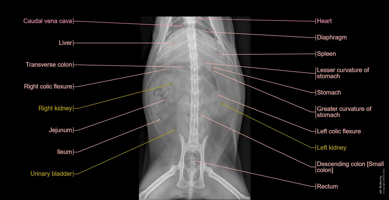
This module of vet-Anatomy is a basic atlas of normal imaging of anatomical feline radiology. The 39 sampled x-ray images of healthy cats were performed by Susanne AEB Boroffka (PhD - dipl. ECVDI, Utrecht, Netherland). Those images were categorized topographically into six chapters (head, vertebral column, thoracic limb, pelvic limb, thorax and abdomen/pelvis).
The anatomical terms were labelled by Antoine Micheau - MD, using different colors to improve the survey and the identification of searched structures on each radiograph.
The translation of the anatomical terms was supervised by Barbara Stockler.
These radiographs of animal anatomy are under the copyright of IMAIOS and can not be freely used.
No content available
