

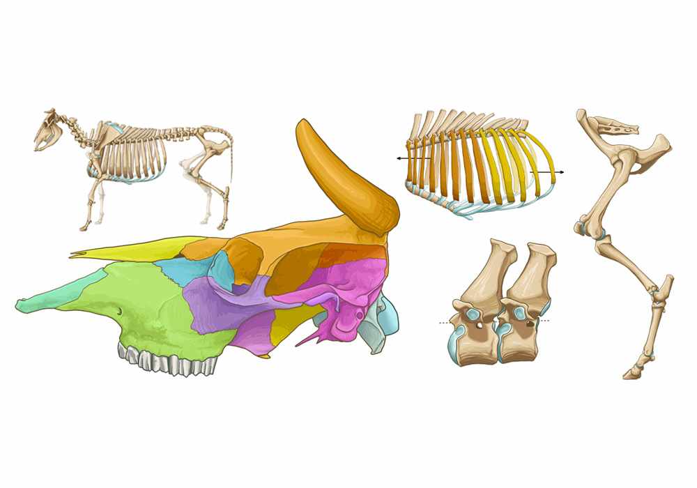
Bovine osteology - Illustrated anatomy atlas of the bones of the bull
Bovine osteology - Illustrated anatomy atlas of the bones of the bull
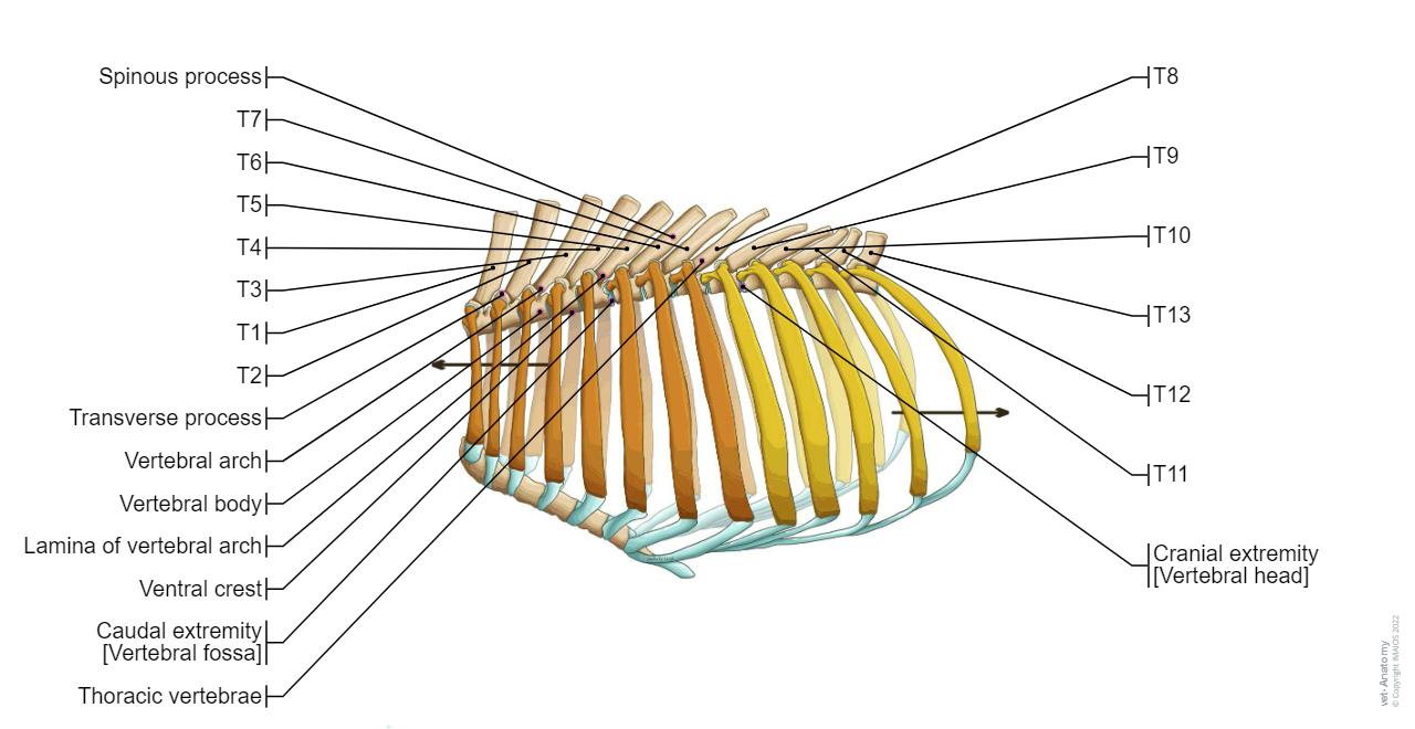
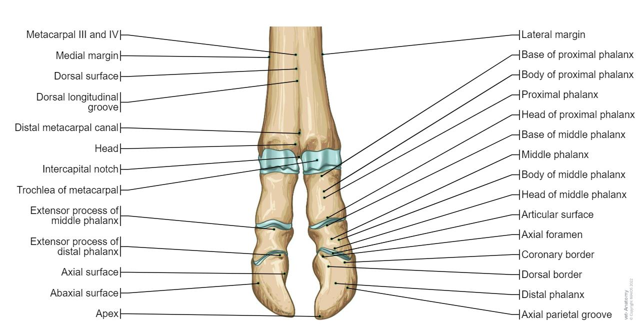
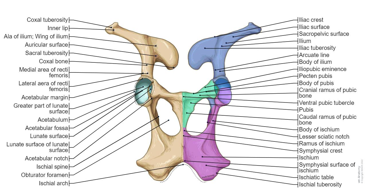
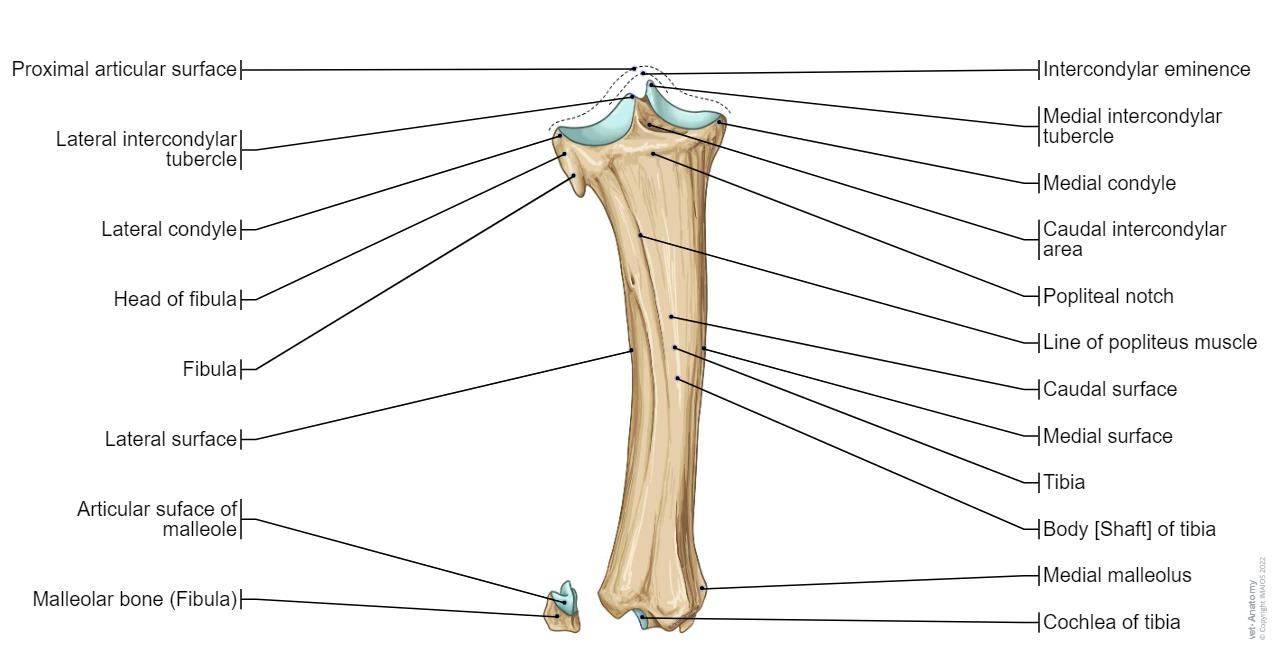
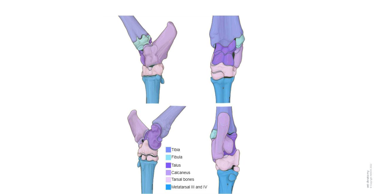
This veterinary anatomy module contains 143 labeled bovine osteology anatomical illustrations. It is particularly useful for veterinary students and rural veterinarians.The main bones of the ox are presented in this anatomical atlas from different anatomical standard points of view, as well as certain joints. The drawings and labeling were done by Estelle Demange, under the anatomical and scientific supervision of Antoine Micheau - MD and Denis Hoa - MD. The translation of the anatomical terms was supervised by Barbara Stockler.
These original illustrations of animal anatomy are under the copyright of IMAIOS and cannot be freely used.
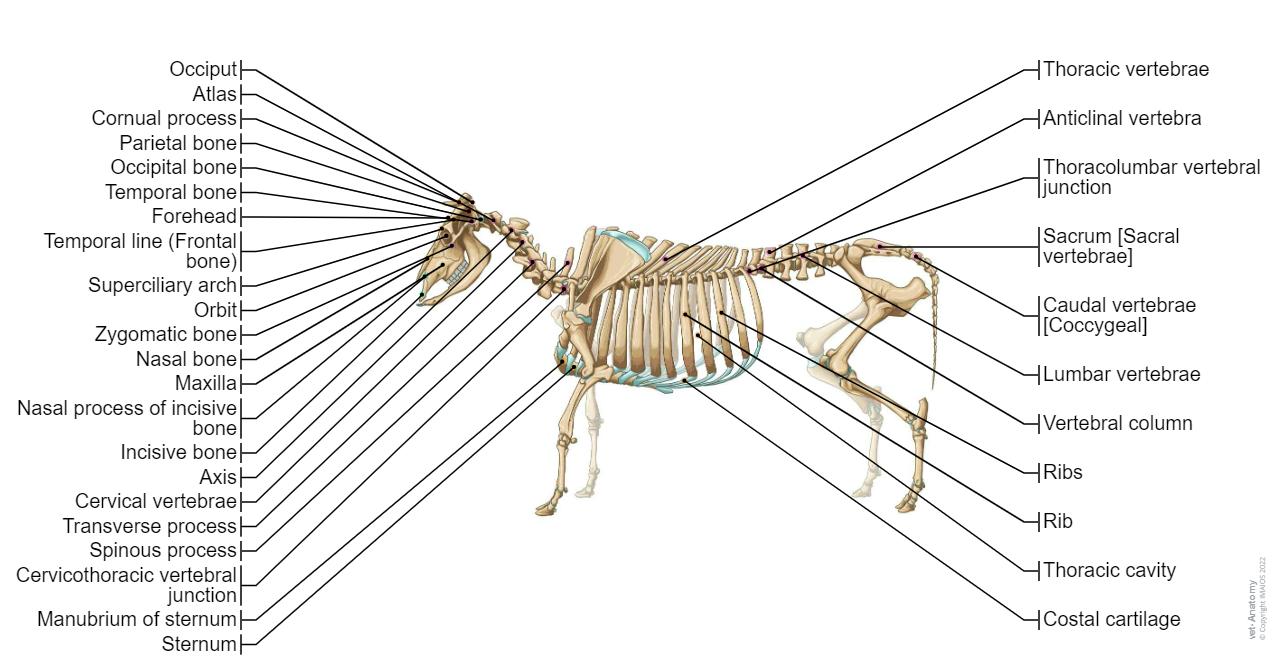

![Sacrum [Sacral vertebrae] – (Bull , Dorsal view)](https://cdn.imaios.cn/i/images/5/6/0/9/469065-9-eng-GB/bull-osteology-illustrations-sacrum.jpg?auto=format,compress&caption=1&q=75&w=1280&s=7e1082f1ad477d475f44d45d12afdda3)

No content available
